Introduction
- Electrophoresis is one of the most important analytical techniques used in biochemistry, molecular biology, and clinical diagnostics.
- It enables the separation of charged molecules such as proteins, nucleic acids, peptides, and lipoproteins based on their movement in an electric field.
- Among the various electrophoretic techniques, Protein Electrophoresis and Polyacrylamide Gel Electrophoresis (PAGE) are widely used for the identification, characterization, and quantification of biomolecules.
- These techniques play a crucial role in disease diagnosis, protein analysis, genetic studies, and biomedical research.
What is Electrophoresis?
Electrophoresis is a laboratory technique used to separate electrically charged molecules by applying an electric field across a supporting medium.
When an electric current passes through a buffer-containing medium, charged molecules migrate toward the electrode carrying the opposite charge:
- Positively charged molecules (cations) move toward the cathode.
- Negatively charged molecules (anions) move toward the anode.
The rate of migration depends on several factors including charge, size, shape, and properties of the supporting medium.
Principle of Electrophoresis
- The fundamental principle of electrophoresis is based on the movement of charged particles under the influence of an electric field.
- Most biological molecules possess ionizable groups that gain or lose protons depending on the pH of the surrounding medium.
- As a result, these molecules acquire positive or negative charges and migrate through a support medium when subjected to an electric field.
The velocity of migration depends on:
- Net electrical charge of the molecule
- Molecular size
- Molecular shape
- Strength of the electric field
- Characteristics of the supporting medium
Molecules with higher charge and smaller size migrate faster than larger molecules with lower charge.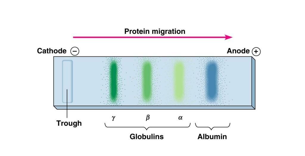
Types of Electrophoresis
- Electrophoresis can be classified based on the medium used, the mechanism of separation, and the purpose of analysis.
- Different types of electrophoresis are employed in clinical laboratories, molecular biology, biotechnology, and research applications.
1. Moving Boundary Electrophoresis
- Moving boundary electrophoresis is the earliest form of electrophoresis, developed by Arne Tiselius.
- In this technique, separation occurs in a free liquid medium without the use of a supporting matrix.
Characteristics
- Separation takes place in a buffer solution.
- Molecules migrate according to their charge and mobility.
- Limited resolving power compared to modern techniques.
- Rarely used in routine laboratories today.
Applications
- Historical studies of serum proteins.
- Research applications.
2. Zone Electrophoresis
- Zone electrophoresis is the most commonly used form of electrophoresis.
- Samples are applied as discrete spots or bands on a supporting medium, where they separate into distinct zones.
Advantages
- High resolution.
- Better separation efficiency.
- Widely used in clinical and research laboratories.
Based on the supporting medium used, zone electrophoresis is further divided into several types.
A. Paper Electrophoresis
- Paper electrophoresis uses filter paper as the supporting medium.
- The paper is saturated with buffer, and samples migrate under the influence of an electric field.
Applications
- Separation of amino acids.
- Separation of small proteins.
- Educational demonstrations.
Limitations
- Lower resolution.
- Time-consuming.
- Largely replaced by modern techniques.
B. Cellulose Acetate Electrophoresis
- In this technique, cellulose acetate membranes are used as the support medium.
Advantages
- Better resolution than paper electrophoresis.
- Rapid separation.
- Easy interpretation of results.
Applications
- Serum protein electrophoresis.
- Hemoglobin electrophoresis.
- Lipoprotein analysis.
C. Gel Electrophoresis
- Gel electrophoresis uses a gel matrix that acts as a molecular sieve, allowing separation based on size and charge.
Types of Gel Electrophoresis
i. Agarose Gel Electrophoresis
- Agarose gel is commonly used for separating DNA and RNA fragments.
Applications
- DNA fingerprinting.
- PCR product analysis.
- Genetic testing.
- Molecular cloning.
Advantages
- Easy gel preparation.
- Suitable for large nucleic acid fragments.
ii. Polyacrylamide Gel Electrophoresis (PAGE)
- PAGE uses a polyacrylamide gel matrix with highly controlled pore size.
Applications
- Protein separation.
- Molecular weight determination.
- Western blotting.
- Proteomic studies.
Advantages
- High resolving power.
- Excellent separation of proteins differing slightly in size.
D. Capillary Electrophoresis
- Capillary electrophoresis separates molecules inside a narrow capillary tube filled with electrolyte solution.
Advantages
- Very high resolution.
- Rapid analysis.
- Minimal sample requirement.
- Automated operation.
Applications
- Clinical diagnostics.
- DNA sequencing.
- Pharmaceutical analysis.
- Forensic investigations.
E. Isoelectric Focusing (IEF)
- Isoelectric focusing separates proteins according to their isoelectric point (pI), the pH at which the protein carries no net charge.
Principle
- Proteins migrate through a pH gradient until they reach the pH corresponding to their pI.
Applications
- Protein characterization.
- Proteomics research.
- Two-dimensional electrophoresis.
F. Immunoelectrophoresis
- Immunoelectrophoresis combines electrophoresis with antigen-antibody reactions to identify specific proteins.
Applications
- Detection of immunoglobulins.
- Diagnosis of immune disorders.
- Investigation of monoclonal gammopathies.
G. SDS-PAGE (Sodium Dodecyl Sulfate-PAGE)
- SDS-PAGE is a specialized form of PAGE in which proteins are treated with SDS detergent.
Principle
- SDS denatures proteins and imparts a uniform negative charge, allowing separation solely on the basis of molecular weight.
Applications
- Protein molecular weight determination.
- Protein purity assessment.
- Research and biotechnology.
H. Two-Dimensional Electrophoresis (2D-PAGE)
Two-dimensional electrophoresis combines:
- Isoelectric Focusing (IEF) – separation based on isoelectric point.
- SDS-PAGE – separation based on molecular weight.
Advantages
- Extremely high resolution.
- Can separate thousands of proteins simultaneously.
Applications
- Proteomics.
- Biomarker discovery.
- Cancer research.
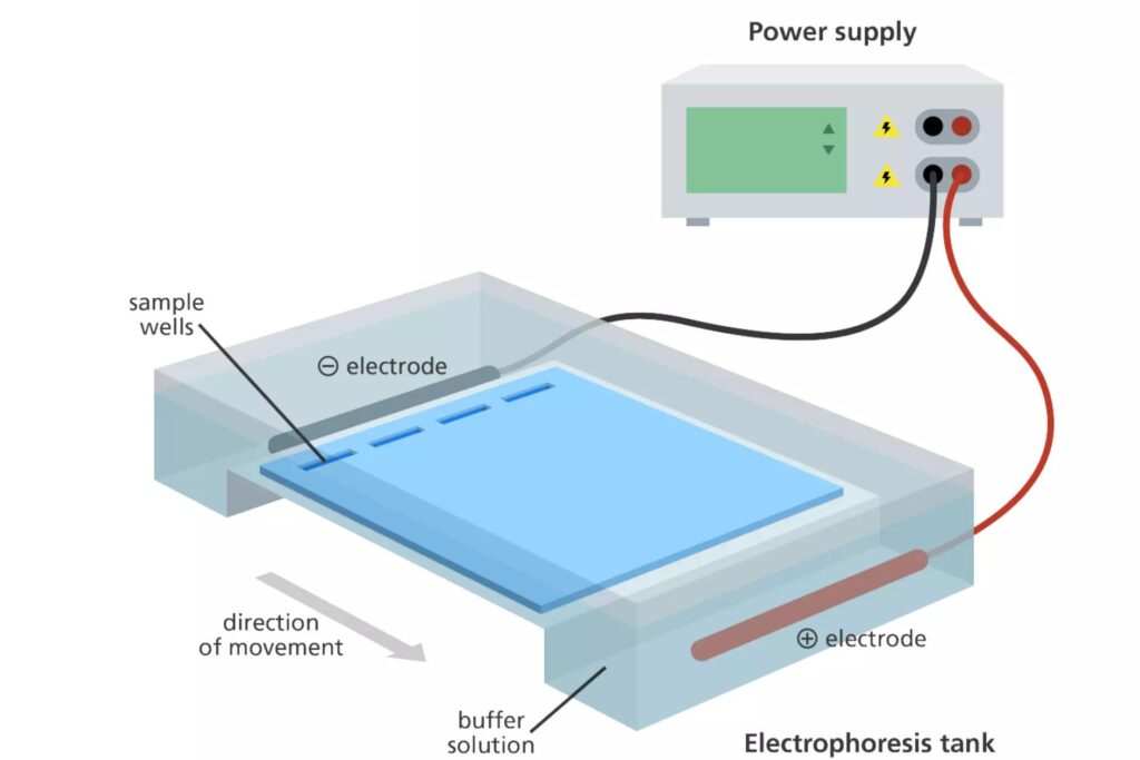
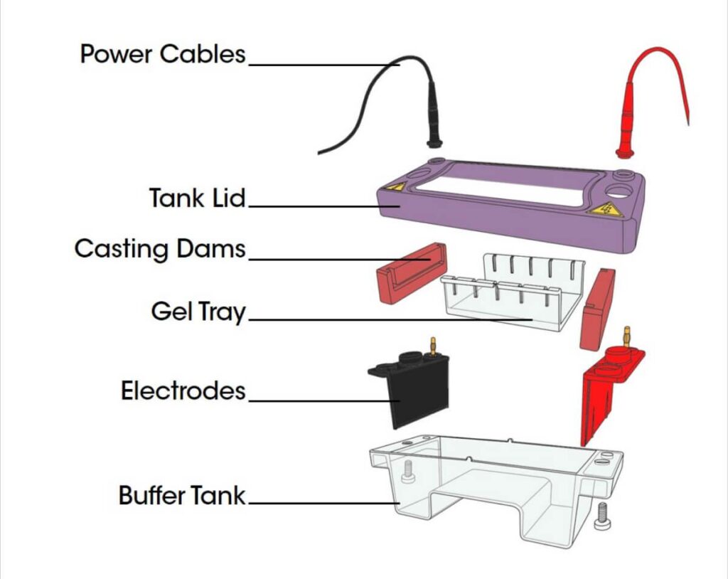
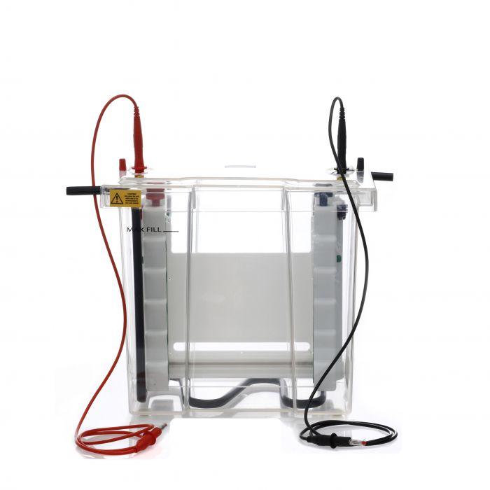
Components
A standard electrophoresis apparatus consists of the following components:
1. Buffer Chambers
- These chambers contain the electrolyte buffer solution that conducts electricity and maintains a constant pH during separation.
2. Electrodes
- Electrodes are usually made of platinum, stainless steel, or carbon.
- Positive electrode = Anode
- Negative electrode = Cathode
They generate the electric field required for migration.
3. Power Supply
- A regulated power supply provides constant voltage or current throughout the experiment.
4. Supporting Medium
- The support medium acts as a matrix through which molecules migrate.
Examples include:
- Filter paper
- Cellulose acetate membrane
- Agarose gel
- Polyacrylamide gel
5. Sample Application System
- A mechanism for loading the biological sample accurately onto the support medium.
6. Protective Cover
- The electrophoresis chamber is covered to prevent evaporation, contamination, and accidental electric shock.
Factors Affecting Electrophoretic Mobility
The movement of molecules during electrophoresis is influenced by several factors.
1. Net Charge – The greater the electrical charge on a molecule, the faster it migrates in an electric field.
2. Molecular Size – Smaller molecules move more rapidly through the support medium than larger molecules.
3. Molecular Shape – Compact molecules migrate faster than elongated or irregularly shaped molecules.
4. Electric Field Strength – Increasing voltage generally increases migration speed, although excessive voltage may generate heat and distort results.
5. Support Medium Characteristics – The pore size and composition of the medium significantly affect molecular movement.
6. Buffer pH – Buffer pH determines the ionization state and net charge of biomolecules.
7. Buffer Ionic Strength – High ionic strength can reduce electrophoretic mobility by increasing resistance.
8. Temperature – Excessive heat can alter protein structure and affect migration patterns.
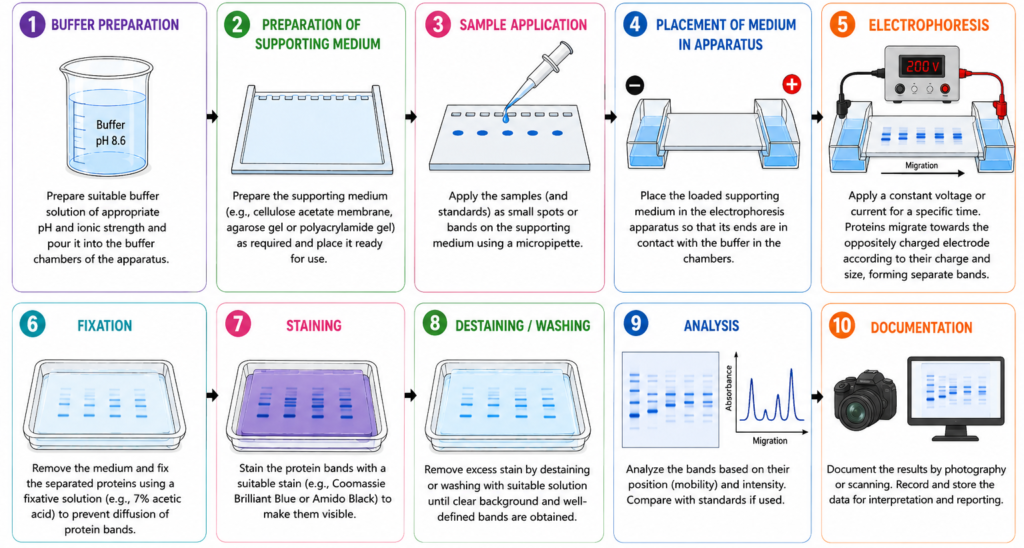
General Procedure
1. Sample Preparation
- The biological sample, such as serum, plasma, urine, or tissue extract, is prepared before analysis.
- Any particulate matter is removed by centrifugation, and the sample may be diluted if required.
- Proper sample preparation ensures accurate and reproducible results.
2. Preparation of the Supporting Medium
A suitable supporting medium is selected depending on the type of electrophoresis being performed. Common media include:
- Cellulose acetate membrane
- Agarose gel
- Polyacrylamide gel (PAGE)
The medium is soaked or prepared with an appropriate buffer solution to maintain optimal pH and conductivity.
3. Sample Application
- A small volume of the prepared sample is carefully applied onto the supporting medium using a micropipette or sample applicator.
- Proper loading is essential to obtain sharp and well-defined protein bands.
4. Electrophoretic Separation
- The supporting medium is placed in the electrophoresis chamber, and the chamber is connected to a power supply.
When an electric field is applied:
- Negatively charged proteins migrate toward the anode.
- Positively charged proteins migrate toward the cathode.
Proteins separate into distinct bands according to their electrophoretic mobility.
5. Fixation of Protein Bands
- After the separation is complete, the medium is treated with a fixative solution to immobilize the proteins and prevent diffusion of the separated bands.
Common fixatives include:
- Acetic acid
- Methanol
- Ethanol-based solutions
6. Staining
- The separated proteins are usually invisible and must be stained for visualization.
Common protein stains include:
- Coomassie Brilliant Blue
- Amido Black
- Bromophenol Blue
- Silver Stain
The stain binds to proteins and produces visible colored bands.
7. Destaining
- Excess stain is removed using a destaining solution.
- This step improves contrast and makes the protein bands more distinct and easier to interpret.
8. Visualization and Documentation
- The stained bands are examined visually or captured using imaging systems.
- The resulting electrophoretic pattern is documented for analysis and record keeping.
9. Quantitative Analysis
- The intensity of protein bands can be measured using a densitometer or computerized image analysis system.
- Quantitative analysis helps determine the relative concentration of different protein fractions.
10. Interpretation of Results
- The final electrophoretic pattern is compared with normal reference patterns.
Variations in the position or intensity of protein bands may indicate specific pathological conditions such as:
- Multiple myeloma
- Liver cirrhosis
- Nephrotic syndrome
- Chronic inflammatory disorders
- Immunoglobulin abnormalities
Serum Protein Electrophoresis
Serum Protein Electrophoresis is a diagnostic technique used to separate serum proteins into distinct fractions.
At alkaline pH (approximately 8.6), serum proteins separate into five major fractions.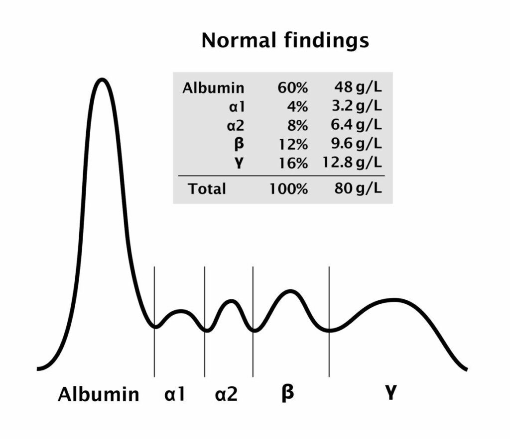
Normal Serum Protein Fractions
| Fraction | Normal Percentage |
|---|---|
| Albumin | 55–65% |
| Alpha-1 Globulin | 2–4% |
| Alpha-2 Globulin | 6–12% |
| Beta Globulin | 8–12% |
| Gamma Globulin | 12–22% |
Albumin migrates fastest toward the anode, while gamma globulins migrate the slowest.
Interpretation
- Serum Protein Electrophoresis (SPEP) is a valuable diagnostic technique used to separate serum proteins into distinct fractions.
- The resulting electrophoretic pattern helps clinicians identify abnormalities associated with various diseases, including liver disorders, kidney diseases, inflammatory conditions, and plasma cell dyscrasias.
Under normal conditions, serum proteins are separated into five major fractions:
- Albumin
- Alpha-1 Globulins
- Alpha-2 Globulins
- Beta Globulins
- Gamma Globulins
Alterations in the concentration or distribution of these fractions can indicate specific pathological conditions.
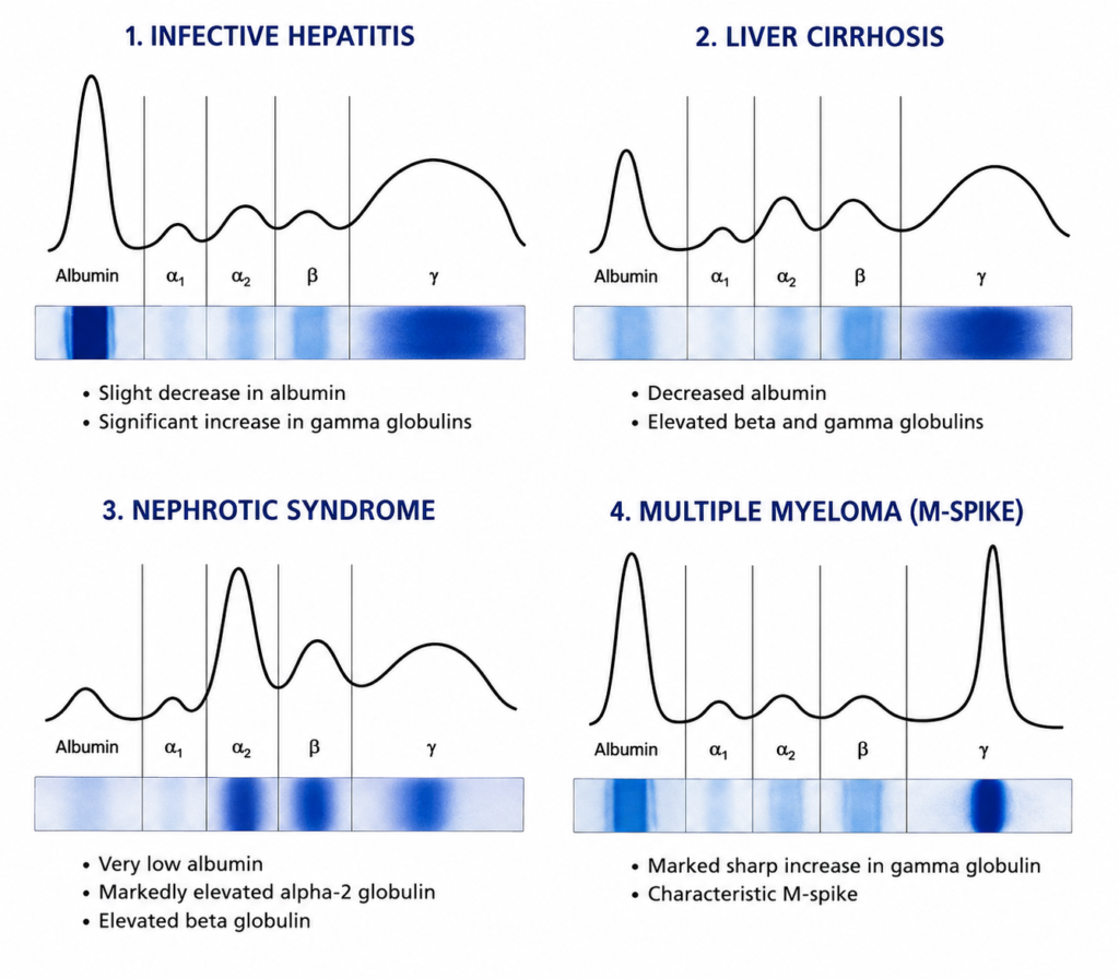
1. Multiple Myeloma
- Multiple myeloma is a malignant proliferation of plasma cells producing excessive monoclonal immunoglobulins.
Electrophoretic Findings
- Sharp, narrow M-spike (Monoclonal Spike) in the gamma region
- Reduced normal immunoglobulin production
- Decreased albumin may be observed
Clinical Significance
The presence of a monoclonal spike strongly suggests:
- Multiple myeloma
- Monoclonal gammopathy of undetermined significance (MGUS)
- Waldenström macroglobulinemia
2. Chronic Inflammatory Disorders
- Chronic infections and inflammatory diseases stimulate increased immunoglobulin production.
Electrophoretic Findings
- Broad increase in gamma globulin region
- Mild increase in alpha-1 and alpha-2 globulins
- Slight reduction in albumin
Clinical Significance
Seen in:
- Tuberculosis
- Rheumatoid arthritis
- Chronic osteomyelitis
- Autoimmune diseases
3. Acute Inflammation
- Acute inflammatory responses increase acute-phase proteins.
Electrophoretic Findings
- Increased alpha-1 globulin fraction
- Increased alpha-2 globulin fraction
- Mild decrease in albumin
- Normal gamma globulin region
Clinical Significance
Observed in:
- Acute bacterial infections
- Trauma
- Burns
- Recent surgery
4. Nephrotic Syndrome
- Nephrotic syndrome causes excessive urinary loss of plasma proteins.
Electrophoretic Findings
- Marked decrease in albumin
- Significant increase in alpha-2 globulin
- Decrease in gamma globulins
- Reduced total serum protein
Clinical Significance
This pattern is characteristic of protein loss through damaged glomeruli.
5. Liver Cirrhosis
- Liver disease impairs albumin synthesis and alters immunoglobulin production.
Electrophoretic Findings
- Decreased albumin
- Increased gamma globulins
- Beta-gamma bridging phenomenon
Clinical Significance
Beta-gamma bridging is a classic feature of hepatic cirrhosis and chronic liver disease.
6. Hypogammaglobulinemia
- This condition results from decreased immunoglobulin production.
Electrophoretic Findings
- Markedly reduced gamma globulin fraction
- Normal or slightly decreased albumin
Clinical Significance
Seen in:
- Primary immunodeficiency disorders
- Congenital agammaglobulinemia
- Immunosuppressive therapy
7. Polyclonal Gammopathy
- Polyclonal gammopathy occurs due to stimulation of multiple plasma cell clones.
Electrophoretic Findings
- Broad diffuse increase in gamma region
- No distinct monoclonal spike
Clinical Significance
Associated with:
- Chronic infections
- Autoimmune diseases
- Chronic liver disorders
8. Protein-Losing Enteropathy
- Excessive loss of proteins occurs through the gastrointestinal tract.
Electrophoretic Findings
- Decreased albumin
- Reduced alpha and gamma globulin fractions
Clinical Significance
Common in:
- Inflammatory bowel disease
- Intestinal lymphangiectasia
- Severe gastrointestinal disorders
Polyacrylamide Gel Electrophoresis
- Polyacrylamide Gel Electrophoresis (PAGE) is a widely used laboratory technique for separating proteins, peptides, and nucleic acids based on their size, charge, and molecular characteristics.
- The technique utilizes a polyacrylamide gel matrix, which acts as a molecular sieve and allows biomolecules to migrate at different rates when an electric field is applied.
- PAGE provides high resolution and excellent separation efficiency, making it one of the most reliable methods for analyzing complex protein mixtures.
- Depending on the experimental requirements, PAGE can be performed as Native PAGE, which preserves the natural structure of proteins, or SDS-PAGE, which separates proteins primarily according to molecular weight.
- This technique is extensively used in clinical diagnostics, molecular biology, biotechnology, proteomics, and biomedical research for protein characterization and purity assessment.
Composition of Polyacrylamide Gel
1. Acrylamide
- Acrylamide is the primary monomer used in gel preparation.
- It polymerizes to form long polyacrylamide chains.
- The concentration of acrylamide determines the pore size of the gel.
- Higher acrylamide concentrations produce smaller pores, while lower concentrations produce larger pores.
2. N,N’-Methylenebisacrylamide (Bis-acrylamide)
- Bis-acrylamide acts as a cross-linking agent.
- It connects polyacrylamide chains to form a stable three-dimensional network.
- The degree of cross-linking affects gel strength and pore size.
3. Buffer Solution
- Buffers maintain a constant pH during electrophoresis.
- Commonly used buffers include Tris-HCl and Tris-Glycine.
- They provide ions necessary for electrical conductivity and protein migration.
4. Ammonium Persulfate (APS)
- APS serves as a polymerization initiator.
- It generates free radicals that initiate the polymerization of acrylamide and bis-acrylamide.
- Fresh APS solution is typically prepared before gel casting.
5. Tetramethylethylenediamine (TEMED)
- TEMED acts as a catalyst for polymerization.
- It accelerates the formation of free radicals from APS.
- This ensures rapid and uniform gel formation.
6. Distilled Water
- Distilled water is used to dissolve all gel components and prepare the desired gel concentration.
- It ensures purity and prevents contamination that could interfere with electrophoresis.
Types of PAGE
1. Native PAGE
Native PAGE separates proteins in their natural or native state without the use of denaturing agents.
Characteristics
- Proteins retain their biological activity and native structure.
- Separation depends on molecular size, shape, and charge.
- Protein-protein interactions remain intact.
Applications
- Analysis of enzyme activity.
- Study of protein complexes.
- Investigation of protein conformations.
2. SDS-PAGE (Sodium Dodecyl Sulfate-PAGE)
SDS-PAGE is the most widely used form of PAGE for protein analysis.
Characteristics
- Proteins are treated with sodium dodecyl sulfate (SDS), a detergent that denatures proteins.
- SDS imparts a uniform negative charge to all proteins.
- Separation occurs primarily according to molecular weight.
Applications
- Determination of protein molecular weight.
- Assessment of protein purity.
- Protein characterization.
- Western blotting.
3. Gradient PAGE
Gradient PAGE utilizes a gel containing gradually increasing concentrations of acrylamide from top to bottom.
Characteristics
- Pore size decreases progressively through the gel.
- Provides improved separation of proteins with a wide range of molecular weights.
- Produces sharper and better-resolved bands.
Applications
- Separation of complex protein mixtures.
- Analysis of proteins with diverse molecular sizes.
- Research and proteomic studies.
4. Two-Dimensional PAGE (2D-PAGE)
Two-dimensional PAGE combines two independent electrophoretic techniques to achieve very high resolution.
First Dimension
- Isoelectric Focusing (IEF)
- Separation based on isoelectric point (pI).
Second Dimension
- SDS-PAGE
- Separation based on molecular weight.
Characteristics
- Separates thousands of proteins simultaneously.
- Provides excellent resolution for complex samples.
Applications
- Proteomics.
- Biomarker discovery.
- Cancer research.
- Comparative protein expression studies.
Principle of PAGE
- In PAGE, proteins migrate through a porous polyacrylamide matrix under the influence of an electric field.
The gel acts as a molecular sieve.
In SDS-PAGE
- All proteins acquire a similar negative charge.
- Separation depends almost entirely on molecular weight.
- Smaller proteins migrate faster.
In Native PAGE
- Separation depends on charge, shape, and molecular size.
Applications of PAGE
1. Protein Separation and Analysis
- PAGE is commonly used to separate proteins based on their molecular size and charge.
- It helps researchers study complex protein mixtures and identify individual protein components.
2. Determination of Molecular Weight
- SDS-PAGE is widely employed to estimate the molecular weight of proteins by comparing their migration with standard protein markers of known sizes.
3. Assessment of Protein Purity
- PAGE helps evaluate the purity of isolated or purified proteins. The presence of a single band indicates high purity, while multiple bands suggest contamination.
4. Protein Characterization
- The technique is used to study protein structure, subunits, isoforms, and post-translational modifications, providing valuable information about protein properties.
5. Western Blotting
- PAGE serves as the initial step in Western blotting. Proteins separated by PAGE are transferred onto a membrane and detected using specific antibodies.
6. Proteomics Research
- Two-Dimensional PAGE (2D-PAGE) is extensively used in proteomics to analyze the expression of thousands of proteins simultaneously and identify disease-related biomarkers.
7. Enzyme Studies
- Native PAGE is used to investigate enzyme activity, protein-protein interactions, and functional protein complexes while preserving their biological activity.
8. Clinical Diagnostics
- PAGE assists in the diagnosis and investigation of various disorders by analyzing abnormal proteins, hemoglobin variants, and disease-associated biomarkers.
9. Genetic and Molecular Biology Research
- PAGE is used for the separation and analysis of DNA and RNA fragments, particularly in DNA sequencing, mutation detection, and gene expression studies.
10. Biotechnology and Pharmaceutical Industry
- The technique is widely employed in the development and quality control of recombinant proteins, vaccines, monoclonal antibodies, and other biopharmaceutical products.
11. Forensic Science
- PAGE is utilized in forensic laboratories for the analysis of biological samples, DNA profiling, and identification of individuals in criminal investigations.
12. Biomarker Discovery
- Researchers use PAGE to identify novel protein biomarkers associated with diseases such as cancer, cardiovascular disorders, diabetes, and neurodegenerative conditions.
Advantages of Polyacrylamide Gel Electrophoresis (PAGE)
Polyacrylamide Gel Electrophoresis (PAGE) is one of the most widely used techniques for the separation and analysis of proteins and nucleic acids due to its high accuracy and resolving power.
1. High Resolution
- PAGE provides excellent separation of molecules that differ only slightly in size or charge, making it highly effective for protein analysis.
2. Adjustable Pore Size
- The pore size of the gel can be controlled by changing the concentration of acrylamide, allowing separation of molecules with different molecular weights.
3. High Sensitivity
- Even small amounts of proteins or nucleic acids can be detected after staining, making PAGE suitable for research and diagnostic purposes.
4. Good Reproducibility
- PAGE produces consistent and reproducible results when performed under standardized conditions.
5. Suitable for Protein Characterization
- The technique helps determine protein purity, molecular weight, subunit composition, and structural variations.
6. Versatile Technique
- PAGE can be performed in different forms such as Native PAGE, SDS-PAGE, Gradient PAGE, and Two-Dimensional PAGE, depending on the analytical requirement.
7. Compatible with Advanced Techniques
- Separated proteins can be further analyzed using Western blotting, mass spectrometry, and proteomic studies.
8. Cost-Effective
- Compared to many advanced analytical techniques, PAGE is relatively inexpensive and widely available in laboratories.
Limitations of Polyacrylamide Gel Electrophoresis (PAGE)
Despite its numerous advantages, PAGE has certain limitations that must be considered during laboratory analysis.
1. Time-Consuming Procedure
- Preparation of gels, electrophoretic separation, staining, and destaining require considerable time.
2. Technical Expertise Required
- Accurate gel preparation and interpretation of results require trained personnel and laboratory experience.
3. Toxic Nature of Acrylamide
- Unpolymerized acrylamide is neurotoxic and must be handled carefully using appropriate safety measures.
4. Limited Sample Capacity
- Only small sample volumes can be loaded onto the gel, restricting large-scale analyses.
5. Protein Denaturation in SDS-PAGE
- Proteins lose their native structure during SDS-PAGE, making it unsuitable for studying biological activity.
6. Difficulty in Recovering Proteins
- Extraction of intact proteins from the gel after separation can be challenging.
7. Heat Generation
- High voltage may generate heat, which can affect protein migration and reduce resolution if not properly controlled.
8. Limited Separation of Very Large Molecules
- Extremely large proteins and nucleic acid fragments may not separate efficiently in polyacrylamide gels.
Clinical Importance of Electrophoresis
Electrophoresis is an essential laboratory technique in clinical biochemistry and diagnostic medicine. It helps identify abnormal proteins, evaluate disease conditions, and monitor treatment outcomes.
1. Diagnosis of Multiple Myeloma
- Serum protein electrophoresis detects monoclonal immunoglobulins (M-proteins), which are characteristic of multiple myeloma and other plasma cell disorders.
2. Evaluation of Liver Diseases
- Changes in albumin and globulin fractions help assess liver cirrhosis, hepatitis, and chronic liver disorders.
3. Detection of Nephrotic Syndrome
- Electrophoretic patterns reveal protein loss through the kidneys, aiding in the diagnosis of nephrotic syndrome.
4. Assessment of Inflammatory and Autoimmune Disorders
- Increases in specific globulin fractions help identify chronic infections, autoimmune diseases, and inflammatory conditions.
5. Identification of Hemoglobin Variants
Hemoglobin electrophoresis is used to diagnose inherited hemoglobin disorders such as:
- Sickle Cell Disease
- Thalassemia
- Hemoglobin C Disease
6. Lipoprotein Analysis
- Electrophoresis helps separate and analyze lipoproteins, which are important in evaluating cardiovascular disease risk.
7. Detection of Immunodeficiency Disorders
- Abnormal gamma globulin levels can indicate congenital or acquired immunodeficiency states.
8. Analysis of Cerebrospinal Fluid (CSF)
- Electrophoresis of CSF proteins assists in diagnosing neurological disorders such as multiple sclerosis.
9. Monitoring Disease Progression
- Electrophoretic patterns can be used to monitor treatment response and disease progression in various chronic conditions.
10. Research and Biomarker Discovery
- Electrophoresis plays a crucial role in identifying novel biomarkers for cancer, cardiovascular diseases, diabetes, and other disorders.