Introduction
- Enzyme-Linked Immunosorbent Assay (ELISA) is one of the most widely used immunological techniques for detecting and measuring antigens or antibodies.
- It is based on the specific binding between an antigen and its corresponding antibody.
- ELISA uses an enzyme-labeled antibody and a color-producing substrate to detect immune reactions.
- The intensity of the color produced is proportional to the amount of antigen or antibody present in the sample.
- ELISA is a highly sensitive, specific, rapid, and cost-effective laboratory technique.
- It is widely used in clinical diagnosis, blood banking, microbiology, immunology, research laboratories, and vaccine development.
- ELISA has largely replaced many older immunological techniques because of its high accuracy, automation, and ability to process large numbers of samples simultaneously.
Principle of ELISA
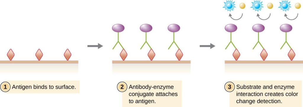
- ELISA is based on the specific interaction between an antigen and its corresponding antibody.
- One of the reactants (antigen or antibody) is immobilized on the surface of a microtiter plate.
- The corresponding antibody or antigen binds specifically to it.
- An enzyme-linked antibody is then added, followed by a suitable substrate.
- The enzyme converts the substrate into a colored product.
- The intensity of the color is measured using an ELISA reader (microplate reader) and is directly proportional to the concentration of the antigen or antibody present in the sample.
Components Required for ELISA
The following components are essential for performing an ELISA test accurately:
| Component | Function |
|---|---|
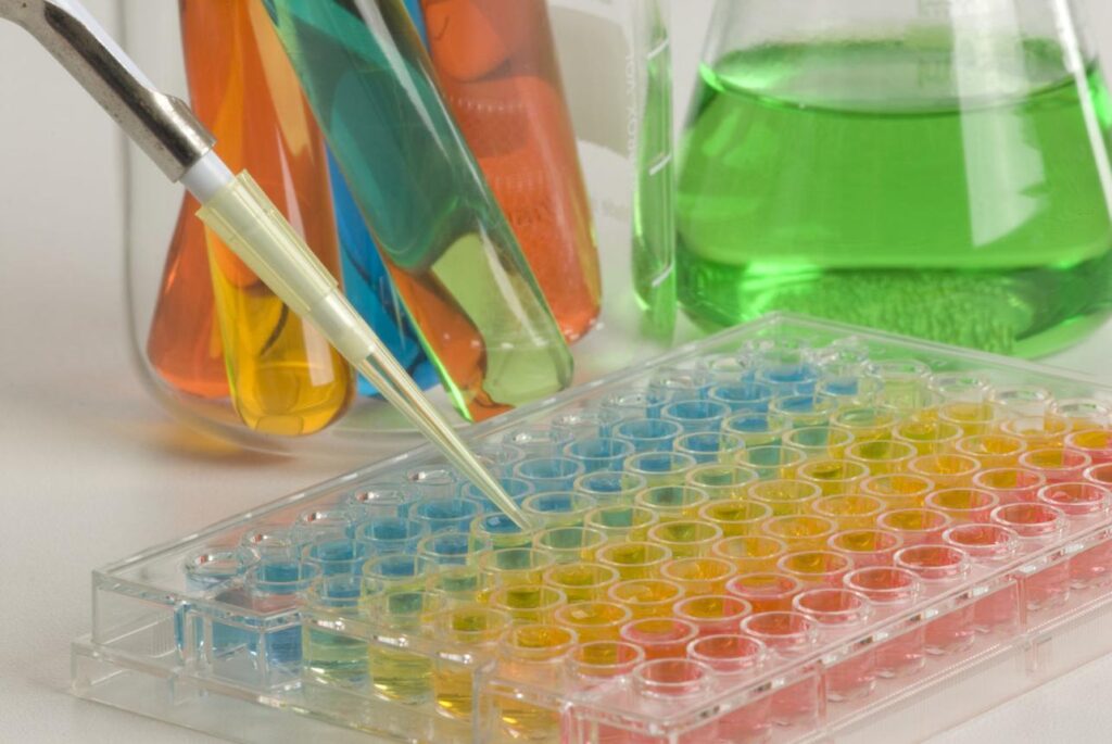
| Microtiter Plate (96-well plate) | Solid surface where the antigen or antibody is immobilized. |
| Antigen | The target molecule to be detected in the sample. |
| Primary Antibody | Specifically binds to the target antigen. |
| Enzyme-Conjugated Secondary Antibody | Binds to the primary antibody and carries an enzyme for color development. |
| Blocking Buffer | Blocks unoccupied binding sites to prevent non-specific binding. |
| Wash Buffer | Removes unbound reagents and reduces background signals. |
| Substrate (e.g., TMB) | Reacts with the enzyme to produce a colored product. |
| Stop Solution | Stops the enzyme reaction and stabilizes the developed color. |
| Sample (Serum, Plasma, Urine, etc.) | Contains the antigen or antibody to be tested. |
| ELISA Reader (Microplate Reader) | Measures the color intensity (optical density) to determine the test result. |
Types of ELISA
ELISA is classified into four main types based on the method used to detect the antigen or antibody.
- Direct ELISA
- Indirect ELISA
- Sandwich ELISA
- Competitive ELISA
Each type has a different principle, procedure, and clinical application.
Direct ELISA
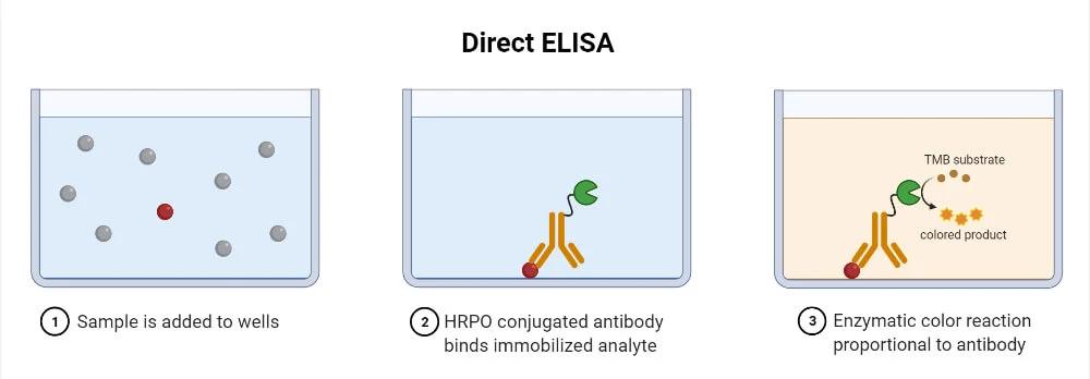
- Direct ELISA is the simplest form of ELISA in which the antigen is directly detected by an enzyme-labeled primary antibody.
- Since only one antibody is used, the procedure is quick and requires fewer steps.
Principle
- The antigen is immobilized on the surface of a microtiter plate.
- An enzyme-conjugated primary antibody is added, which binds specifically to the antigen. After washing, a suitable substrate is added.
- The enzyme converts the substrate into a colored product.
- The intensity of the color is directly proportional to the amount of antigen present.
Procedure
- Coat plate: Add sample directly to the well — antigen adsorbs non-specifically to the polystyrene surface. Incubate at 4°C overnight or 37°C for 2 hours.
- Block: Add blocking buffer (1–5% BSA or skimmed milk in PBS) for 1 hour at 37°C to prevent non-specific antibody binding to bare polystyrene.
- Wash: 3–5 times with PBS-Tween wash buffer.
- Add enzyme-conjugated primary antibody: Specific for the target antigen; labelled directly with HRP or alkaline phosphatase. Incubate 1–2 hours.
- Wash: Remove unbound conjugated antibody — 3–5 washes.
- Add substrate: TMB (for HRP) or PNPP (for AP). Incubate 15–30 minutes in the dark.
- Stop reaction: Add stop solution (H₂SO₄ for TMB; NaOH for PNPP).
- Read absorbance: Measure at 450 nm (TMB) or 405 nm (PNPP) using an ELISA reader.
Applications
- Detection of microbial antigens.
- Detection of viral proteins.
- Quality control in research laboratories.
- Measurement of specific proteins.
Advantages
- Simple and rapid.
- Fewer reagents required.
- Short incubation time.
- Lower risk of cross-reactivity.
Limitations
- Lower sensitivity than indirect ELISA.
- Each primary antibody must be enzyme-labeled.
- Less signal amplification.
Indirect ELISA
- Indirect ELISA is commonly used to detect antibodies in patient serum.
- It is one of the most widely used ELISA techniques because of its high sensitivity and flexibility.
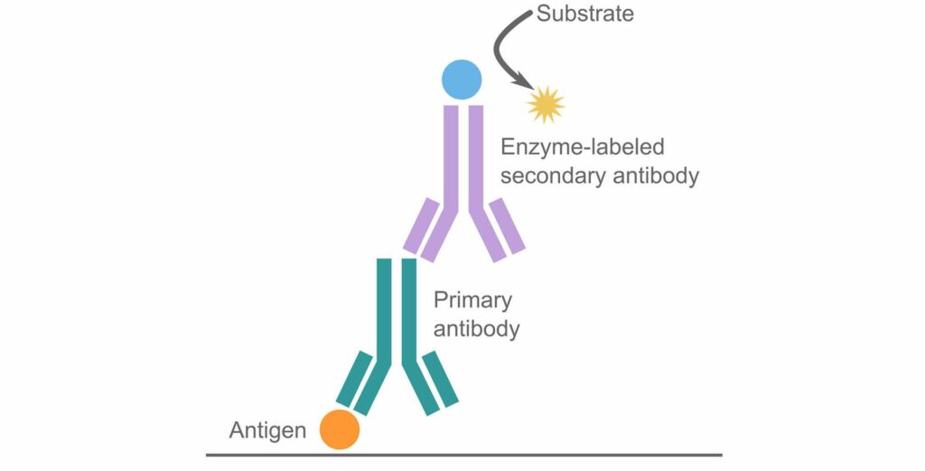
Principle
- The antigen is first coated onto the microtiter plate.
- The patient’s serum is added, allowing specific antibodies (if present) to bind to the antigen.
- An enzyme-conjugated secondary antibody is then added, which binds to the primary antibody.
- After adding the substrate, a colored reaction develops.
- The color intensity is directly proportional to the amount of antibody present in the sample.
Procedure
- Coat plate with known antigen: Add antigen solution (specific concentration per kit instructions). Incubate at 4°C overnight.
- Block: 1–5% BSA or milk for 1 hour at 37°C.
- Wash: 3–5 times with PBS-Tween.
- Add patient serum (primary antibody): Diluted as per kit instructions (typically 1:100 to 1:400). Incubate 1–2 hours at 37°C.
- Wash: Remove unbound patient antibodies — 3–5 washes.
- Add enzyme-conjugated secondary antibody: Anti-human IgG (or IgM) labelled with HRP or alkaline phosphatase. Incubate 1 hour at 37°C.
- Wash: Remove unbound secondary antibody.
- Add substrate: TMB or PNPP. Incubate 15–30 minutes in dark.
- Stop reaction and read absorbance.
Interpretation:
- Optical Density (OD) above cut-off value = Reactive (antibodies detected)
- OD below cut-off = Non-reactive
- The cut-off is determined by the mean OD of negative controls plus 2–3 standard deviations, as specified in the kit insert
Applications
- Detection of HIV antibodies.
- Diagnosis of Hepatitis B and Hepatitis C infections.
- Detection of COVID-19 antibodies.
- Diagnosis of Dengue, Typhoid, and other infectious diseases.
- Detection of autoantibodies in autoimmune disorders.
- Blood donor screening.
Advantages
- Highly sensitive and specific.
- Detects very low concentrations of antibodies.
- One enzyme-conjugated secondary antibody can be used for different primary antibodies.
- Suitable for testing a large number of samples.
- Cost-effective and widely used in diagnostic laboratories.
Limitations
- Requires two antibodies, increasing the number of steps.
- Longer procedure than Direct ELISA.
- Possibility of non-specific binding if washing is inadequate.
- Cross-reactivity may occur with closely related antibodies.
Sandwich ELISA
- Sandwich ELISA is one of the most sensitive and specific ELISA techniques.
- It is mainly used to detect and quantify antigens present in biological samples.
- In this method, the antigen is “sandwiched” between two specific antibodies: a capture antibody and a detection antibody.
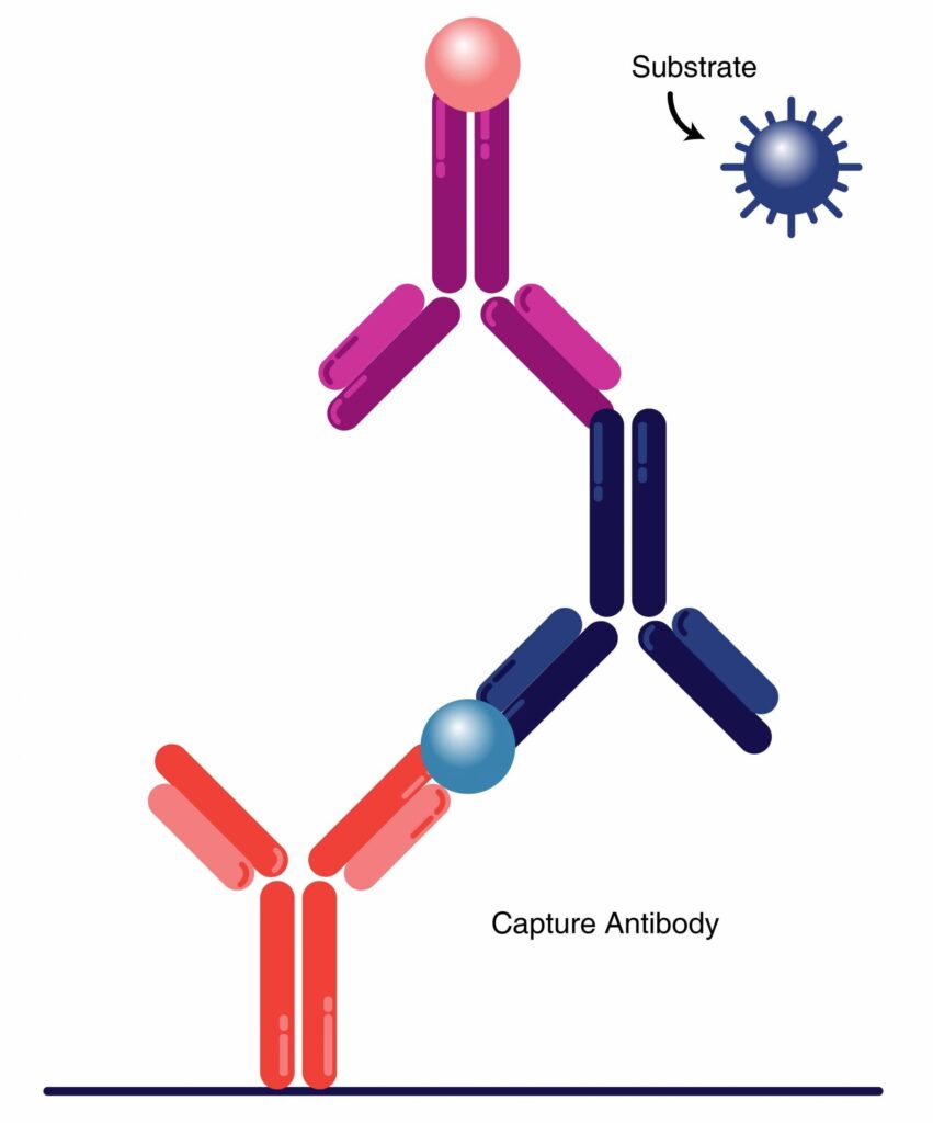
Principle
- A capture antibody is first coated onto the microtiter plate.
- The sample containing the antigen is added, allowing the antigen to bind to the capture antibody.
- Next, an enzyme-conjugated detection antibody binds to another epitope on the antigen, forming an antibody–antigen–antibody complex.
- After adding the substrate, a colored product is formed.
- The color intensity is directly proportional to the concentration of the antigen.
Procedure
Step 1: Coat the Microtiter Plate
The wells of a 96-well microtiter plate are coated with a capture antibody specific to the target antigen.
Step 2: Blocking
A blocking buffer is added to block the remaining unoccupied sites and prevent non-specific binding.
Step 3: Add the Sample
The patient sample containing the antigen is added to the wells and incubated. The antigen binds specifically to the capture antibody.
Step 4: Wash the Plate
The wells are washed with wash buffer to remove any unbound substances.
Step 5: Add the Detection Antibody
A detection antibody, which binds to a different epitope of the antigen, is added and incubated.
Step 6: Wash the Plate
Wash the wells again to remove any unbound detection antibody.
Step 7: Add the Enzyme-Conjugated Antibody
An enzyme-conjugated secondary antibody is added (if the detection antibody is not already enzyme-labeled). It binds to the detection antibody.
Step 8: Wash the Plate
Wash the wells thoroughly to remove any excess enzyme-conjugated antibody.
Step 9: Add the Substrate
A suitable chromogenic substrate (such as TMB) is added. The enzyme reacts with the substrate to produce a colored product.
Step 10: Stop the Reaction
Add a stop solution (usually sulfuric acid) to stop the enzyme reaction and stabilize the color.
Step 11: Read the Results
Measure the optical density (OD) using an ELISA reader (microplate reader). The color intensity is directly proportional to the concentration of the antigen in the sample.
Applications
- Measurement of cytokines (e.g., IL-6, TNF-α).
- Detection of hormones (e.g., insulin, TSH).
- Estimation of tumor markers.
- Detection of viral and bacterial antigens.
- Quantification of proteins in research laboratories.
Advantages
- Very high sensitivity and specificity.
- Suitable for complex biological samples.
- Accurate quantitative measurement.
- Low background interference.
Limitations
- More expensive than Direct and Indirect ELISA.
- Requires two antibodies that recognize different epitopes.
- Procedure is longer and more complex.
Competitive ELISA
- Competitive ELISA is used to measure the concentration of small antigens, hormones, drugs, toxins, and other molecules that have only one antigenic epitope.
- In this method, the sample antigen competes with a labeled antigen for binding to a limited number of antibody binding sites.
- Unlike other ELISA methods, the color intensity is inversely proportional to the concentration of the antigen in the sample.
Principle
- A specific antibody is incubated with the sample containing the antigen.
- The mixture is then added to an antigen-coated microtiter plate.
- Any free antibody binds to the coated antigen.
- After washing, an enzyme-conjugated secondary antibody and substrate are added.
- The amount of color produced is inversely related to the amount of antigen present in the sample.
Higher antigen concentration → Less color development
Lower antigen concentration → More color development
Procedure
- Coat the microtiter plate with a known antigen.
- Mix the sample antigen with a specific primary antibody.
- Add the mixture to the antigen-coated wells.
- Incubate to allow competition for antibody binding.
- Wash the wells to remove unbound materials.
- Add the enzyme-conjugated secondary antibody.
- Wash the wells again.
- Add the substrate solution and allow color to develop.
- Add the stop solution.
- Measure the absorbance using an ELISA reader.
Applications
- Measurement of steroid hormones (e.g., cortisol and progesterone).
- Detection of drugs and drug abuse screening.
- Detection of toxins and pesticides.
- Measurement of small proteins and peptides.
- Therapeutic drug monitoring.
Advantages
- Highly sensitive for detecting small molecules.
- Suitable for antigens with a single epitope.
- Can measure low concentrations of analytes.
- Useful for hormone and drug assays.
Limitations
- More complex than other ELISA methods.
- Interpretation of results can be difficult.
- Color intensity is inversely proportional to antigen concentration.
- Requires careful optimization of reagents.
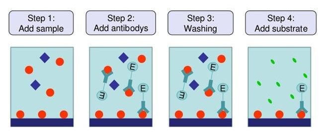
General ELISA Procedure
Although the procedure varies slightly depending on the type of ELISA, the basic steps are similar.
1. Coating – The antigen or antibody is coated onto the wells of a 96-well microtiter plate and allowed to bind to the surface.
2. Blocking – A blocking buffer is added to cover the remaining unoccupied sites and prevent non-specific binding.
3. Sample Addition – The patient sample containing the antigen or antibody is added to the wells and incubated to allow specific binding.
4. Washing – The wells are washed with wash buffer to remove any unbound substances.
5. Addition of Enzyme-Conjugated Antibody – An enzyme-linked antibody is added, which binds specifically to the target antigen or antibody.
6. Second Washing – The wells are washed again to remove any excess enzyme-conjugated antibody.
7. Substrate Addition – A suitable chromogenic substrate (such as TMB) is added. The enzyme reacts with the substrate to produce a colored product.
8. Stop Reaction – A stop solution (usually sulfuric acid) is added to stop the enzyme reaction and stabilize the color.
9. Reading the Results – The color intensity is measured using an ELISA reader (microplate reader). The optical density (OD) is directly proportional to the concentration of the target analyte (except in Competitive ELISA).
Interpretation of ELISA Results
The ELISA reader measures the optical density (OD) of each well. The OD value indicates the amount of antigen or antibody present in the sample.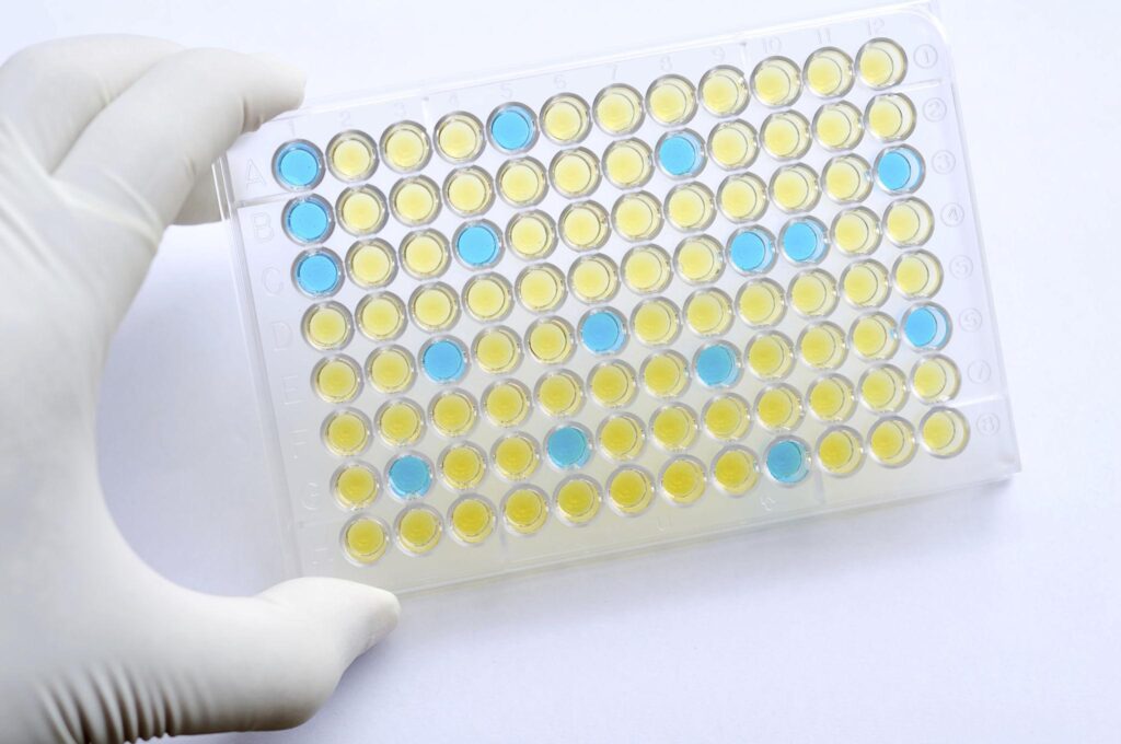
| Result | Interpretation |
|---|---|
| High OD value | High concentration of antigen or antibody (except Competitive ELISA). |
| Low OD value | Low concentration of antigen or antibody. |
| No color development | Negative result or absence of the target analyte. |
| Competitive ELISA | High antigen concentration produces less color, while low antigen concentration produces more color. |
The results are compared with positive controls, negative controls, and standard calibration curves to determine the final interpretation.
Clinical Applications
ELISA is one of the most widely used immunological techniques in clinical laboratories because of its high sensitivity, specificity, and ability to detect a wide range of antigens and antibodies.
1. Diagnosis of Infectious Diseases
ELISA is commonly used to detect antigens or antibodies associated with infectious diseases such as:
- HIV infection
- Hepatitis B (HBV)
- Hepatitis C (HCV)
- Dengue fever
- COVID-19
- Typhoid fever
- Lyme disease
2. Blood Bank Screening
- Screening donated blood for HIV, HBV, HCV, and other transfusion-transmitted infections.
- Ensures the safety of blood and blood products.
3. Hormone Estimation
ELISA is used to measure various hormones, including:
- Insulin
- Thyroid-stimulating hormone (TSH)
- Human chorionic gonadotropin (hCG)
- Cortisol
- Progesterone
- Estrogen
4. Detection of Tumor Markers
ELISA helps detect and monitor tumor markers such as:
- Prostate-specific antigen (PSA)
- Alpha-fetoprotein (AFP)
- Carcinoembryonic antigen (CEA)
- CA-125
- CA 19-9
5. Diagnosis of Autoimmune Diseases
ELISA is used to detect autoantibodies in diseases such as:
- Systemic lupus erythematosus (SLE)
- Rheumatoid arthritis
- Autoimmune thyroid diseases
- Celiac disease
6. Allergy Testing
- Detection of allergen-specific IgE antibodies.
- Identification of food, drug, and environmental allergies.
7. Measurement of Cytokines and Biomarkers
ELISA is widely used to estimate:
- Interleukins (IL-6, IL-1β)
- Tumor Necrosis Factor-alpha (TNF-α)
- C-reactive protein (CRP)
- Procalcitonin (PCT)
- Other inflammatory biomarkers
8. Vaccine Development and Evaluation
- Assessment of antibody response after vaccination.
- Evaluation of vaccine efficacy in clinical trials.
9. Research Applications
- Quantification of proteins and peptides.
- Measurement of growth factors.
- Detection of enzymes and receptors.
- Biomedical and pharmaceutical research.
10. Food and Environmental Testing
- Detection of food allergens.
- Identification of bacterial toxins.
- Monitoring pesticide residues and environmental contaminants.
Advantages
- Highly sensitive and specific.
- Can detect very low concentrations of antigens or antibodies.
- Rapid and easy to perform.
- Suitable for testing a large number of samples simultaneously.
- Quantitative as well as qualitative analysis is possible.
- Easily automated for routine laboratory use.
- Cost-effective compared to many molecular techniques.
- Requires only a small sample volume.
- Widely used in diagnostic, research, and industrial laboratories.
- Produces objective and reproducible results.
Limitations
- False-positive and false-negative results may occur.
- Requires high-quality antibodies and reagents.
- Cross-reactivity may interfere with accuracy.
- Improper washing can increase background signals.
- Enzyme activity may be affected by temperature and pH.
- Cannot always distinguish between current and past infections.
- Requires specialized equipment such as an ELISA reader.
- More time-consuming than rapid diagnostic tests.
Comparison of Different Types of ELISA
| Feature | Direct ELISA | Indirect ELISA | Sandwich ELISA | Competitive ELISA |
|---|---|---|---|---|
| Target Detected | Antigen | Antibody | Antigen | Antigen |
| Number of Antibodies | One | Two | Two | One or Two |
| Sensitivity | Low | High | Very High | High |
| Specificity | Moderate | High | Very High | High |
| Procedure | Simple | Moderate | Complex | Complex |
| Signal Amplification | No | Yes | Yes | No |
| Quantitative | Yes | Yes | Yes | Yes |
| Common Applications | Protein detection | Antibody detection | Hormones, cytokines, tumor markers | Small molecules, drugs, hormones |
ELISA vs Rapid Test vs Western Blot
| Feature | ELISA | Rapid Test | Western Blot |
| Sensitivity | High | Moderate | Very High |
| Specificity | High | Moderate | Very High |
| Time Required | 2–5 hours | 10–30 minutes | Several hours |
| Quantitative | Yes | Usually No | Semi-quantitative |
| Automation | Yes | No | Limited |
| Routine Laboratory Use | Yes | Yes | Mainly Confirmatory |