Introduction
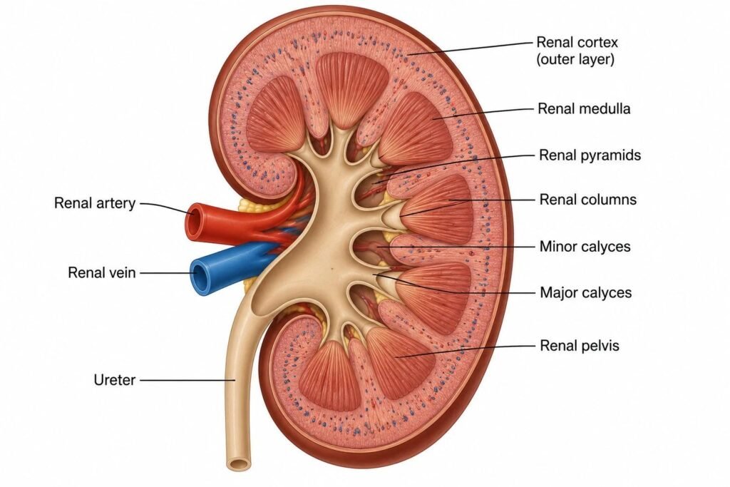
- The kidneys are vital organs that maintain the body’s internal environment by filtering blood, removing metabolic waste products, and regulating fluid, electrolyte, and acid–base balance.
- The kidneys also help regulate blood pressure, produce erythropoietin for red blood cell formation, and activate vitamin D for calcium and phosphate metabolism.
- Renal Function Tests (RFTs) are a group of laboratory investigations performed on blood and urine samples to assess how effectively the kidneys function.
- The major investigations include serum creatinine, blood urea, estimated glomerular filtration rate (eGFR), serum electrolytes, urine albumin-to-creatinine ratio (UACR), and routine urinalysis.
- These tests help detect kidney dysfunction, assess the severity of renal disease, monitor disease progression, and evaluate the response to treatment.
- RFTs help identify abnormalities in glomerular filtration, kidney damage, tubular function, electrolyte balance, and acid–base regulation.
- RFTs are particularly important in patients with diabetes mellitus, hypertension, cardiovascular disease, and other conditions that increase the risk of kidney damage.
- Early detection of kidney dysfunction through appropriate laboratory investigations can help guide treatment and prevent or delay the progression of chronic kidney disease (CKD).

Functions of the Kidneys
- Understanding the physiological functions of the kidneys helps explain the clinical significance of renal function tests.
The major functions of the kidneys include:
1. Excretion of metabolic waste products:
- The kidneys remove waste products such as urea, creatinine, and uric acid from the blood.
2. Regulation of water balance:
- The kidneys regulate the amount of water excreted in urine and help maintain normal body fluid volume.
3. Electrolyte regulation:
- They maintain appropriate concentrations of sodium, potassium, chloride, calcium, and phosphate.
4. Maintenance of acid–base balance:
- The kidneys regulate hydrogen ion excretion and bicarbonate reabsorption to maintain blood pH.
5. Regulation of blood pressure:
- The kidneys help regulate blood pressure through sodium balance and the renin–angiotensin–aldosterone system.
6. Erythropoietin production:
- The kidneys produce erythropoietin, which stimulates red blood cell production in the bone marrow.
7. Vitamin D activation:
- The kidneys convert 25-hydroxyvitamin D into its active form, 1,25-dihydroxyvitamin D, which contributes to calcium and phosphate homeostasis.
Damage to the kidneys may impair one or more of these functions, resulting in biochemical abnormalities that can be detected through laboratory investigations.
Types of Renal Function Tests
The major investigations used to assess renal function are summarized in the following table.
| Test | Sample | Main Purpose |
|---|---|---|
| Serum creatinine | Blood | Evaluates kidney filtration indirectly |
| Blood urea | Blood | Assesses nitrogenous waste accumulation |
| Blood urea nitrogen (BUN) | Blood | Measures nitrogen derived from urea |
| Estimated glomerular filtration rate (eGFR) | Calculated from blood measurements | Estimates the kidney’s filtration capacity |
| Urine albumin-to-creatinine ratio (UACR) | Spot urine | Detects albuminuria and kidney damage |
| Routine urinalysis | Urine | Detects protein, blood, glucose, and other abnormalities |
| Serum electrolytes | Blood | Assesses electrolyte balance |
| Serum bicarbonate | Blood | Helps assess acid–base disturbances |
| Serum uric acid | Blood | Evaluates uric acid concentration |
| Creatinine clearance | Blood and timed urine | Estimates creatinine clearance |
| Urine osmolality | Urine | Assesses urine concentration |
| Fractional excretion of electrolytes | Blood and urine | Helps evaluate renal tubular handling of electrolytes |
The selection of tests depends on the clinical condition and the specific aspect of kidney function being investigated.
Specimen Collection
- Proper specimen collection, handling, and transportation are essential for obtaining accurate and reliable results in renal function testing.
- Blood and urine samples are commonly used to evaluate kidney function.
1. Blood Sample Collection
- Specimen: Serum or plasma.
- Collection method: Blood is collected by venipuncture using standard aseptic techniques.
- Tests performed: Serum creatinine, blood urea, serum electrolytes, uric acid, and other biochemical parameters.
- Sample handling: The sample is processed according to the laboratory’s requirements. Serum is separated after clotting and centrifugation when applicable.
- Precautions: Avoid hemolysis and ensure proper sample identification and transportation.
2. Urine Sample Collection
- Specimen: Fresh, clean-catch midstream urine.
- Collection method: The patient collects urine in a clean, appropriate specimen container after following the recommended cleaning procedure.
- Tests performed: Routine urinalysis, urine protein estimation, and urine albumin-to-creatinine ratio (UACR).
- Sample handling: The sample should be transported promptly to the laboratory and analyzed as soon as possible.
- Precautions: Avoid contamination with stool, water, or other foreign substances.
3. Twenty-Four-Hour Urine Collection
- Specimen: Twenty-four-hour urine sample.
- Collection method: The patient discards the first urine passed at the beginning of the collection period, records the starting time, and collects all subsequent urine for the next 24 hours, including the final urine passed at the designated end time.
- Tests performed: Creatinine clearance and other selected urinary investigations.
- Sample handling: The total urine volume is measured and recorded, and the sample is mixed thoroughly before the required aliquot is sent for analysis.
- Precautions: The entire urine output must be collected. Missing even one collection can affect the accuracy of the results.
4. Patient Preparation
- Follow the laboratory’s instructions regarding fasting and fluid intake.
- Inform the healthcare professional about medications and supplements that may influence test results.
- Avoid strenuous exercise before urine albumin testing when advised.
- Do not discontinue prescribed medications without medical advice.
5. Sample Labelling and Transportation
- Label each specimen with the patient’s identification details, collection date, and collection time, as applicable.
- Use appropriate, leak-proof specimen containers.
- Transport samples promptly to the laboratory.
- Follow the laboratory’s recommended storage and processing conditions if immediate analysis is not possible.
Serum Creatinine
- Serum creatinine is one of the most commonly measured biochemical parameters in renal function testing.
- Creatinine is produced mainly from the metabolism of creatine and phosphocreatine in skeletal muscle.
- It enters the bloodstream at a relatively steady rate and is removed predominantly through glomerular filtration.
- When kidney filtration decreases, serum creatinine generally increases.
Principle
- Serum creatinine concentration reflects the balance between creatinine production and its elimination by the kidneys.
- An increase in serum creatinine may indicate reduced glomerular filtration, although the result is also influenced by muscle mass, diet, medications, and other factors.
Methods of Estimation
Serum creatinine can be measured using the following methods:
- Jaffe method:
- This method is based on the reaction between creatinine and alkaline picrate, producing an orange-red complex.
- Certain substances, including glucose, ketones, and some medications, can interfere with the reaction.
- Enzymatic method:
- This method uses enzyme-based reactions to quantify creatinine.
- It is generally more specific than the traditional Jaffe method, although interference may still occur.
- Modern clinical biochemistry laboratories commonly use automated analyzers to measure serum creatinine.
Reference Values
Typical adult reference intervals are approximately:
| Parameter | Approximate Reference Interval |
|---|---|
| Serum creatinine in adult males | 0.7–1.3 mg/dL |
| Serum creatinine in adult females | 0.6–1.1 mg/dL |
These values are illustrative rather than universal. The reference interval varies with the laboratory method, population, age, sex, and muscle mass.
Clinical Significance
Increased serum creatinine may be observed in:
- Acute kidney injury.
- Chronic kidney disease.
- Reduced renal perfusion.
- Urinary tract obstruction.
- Severe dehydration.
- Certain medication-related conditions.
Blood Urea and Blood Urea Nitrogen
- Urea is the major nitrogen-containing waste product formed during amino acid and protein metabolism.
- When amino acids are broken down, ammonia is produced.
- Because ammonia is toxic, the liver converts it into urea through the urea cycle.
- Urea enters the bloodstream and is subsequently excreted by the kidneys.
- When renal excretion decreases, blood urea concentrations may increase.
Difference Between Blood Urea and BUN
Blood urea and blood urea nitrogen measure related but different quantities.
- Blood urea: Measures the concentration of the entire urea molecule.
- Blood urea nitrogen (BUN): Measures only the nitrogen component of urea.
Consequently, their numerical values are not interchangeable.
Reference Values
| Parameter | Approximate Adult Reference Interval |
|---|---|
| Blood urea | 15–40 mg/dL |
| BUN | 7–20 mg/dL |
Reference intervals vary between laboratories and populations.
Clinical Significance
Increased blood urea may occur in:
- Reduced kidney function.
- Dehydration.
- Gastrointestinal bleeding.
- Increased protein intake.
- Increased protein catabolism.
- Reduced renal blood flow.
Decreased blood urea may be observed in severe liver dysfunction, low protein intake, or certain states of overhydration.
Unlike creatinine, urea is strongly influenced by dietary protein intake, liver function, hydration, and catabolic activity. Therefore, blood urea alone is not a reliable measure of glomerular filtration.
Estimated Glomerular Filtration Rate
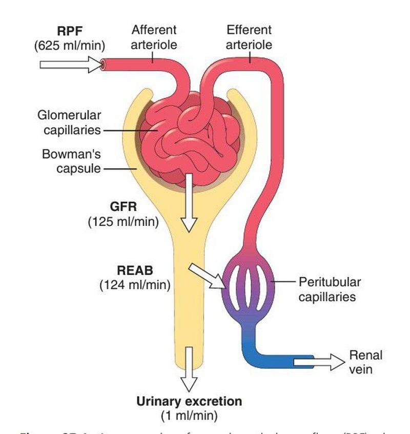
- The glomerular filtration rate (GFR) is the volume of plasma filtered by the glomeruli per unit of time.
- It is one of the most important indicators of kidney filtration function.
- The estimated glomerular filtration rate (eGFR) is calculated using equations that incorporate serum creatinine and, depending on the equation, variables such as age and sex.
- Validated equations using cystatin C, either alone or combined with creatinine, may also be used when a more accurate estimate is needed.
Clinical Significance
The eGFR helps clinicians:
- Assess kidney filtration.
- Identify and classify chronic kidney disease.
- Monitor changes in renal function.
- Evaluate patients at risk of kidney disease.
- Guide medication dosing when appropriate.
eGFR Categories
The following categories are used in the classification of chronic kidney disease.
| GFR Category | eGFR (mL/min/1.73 m²) | Interpretation |
|---|---|---|
| G1 | ≥90 | Normal or high |
| G2 | 60–89 | Mildly decreased |
| G3a | 45–59 | Mildly to moderately decreased |
| G3b | 30–44 | Moderately to severely decreased |
| G4 | 15–29 | Severely decreased |
| G5 | <15 | Kidney failure range |
Important: An eGFR of 60–89 mL/min/1.73 m² does not, by itself, establish chronic kidney disease. Similarly, an eGFR of 90 or above does not exclude kidney damage.
- For a diagnosis of CKD, abnormalities of kidney structure or function generally need to persist for at least three months.
- Evidence of kidney damage, such as persistent albuminuria, can establish CKD even when eGFR is relatively preserved.
Urine Albumin-to-Creatinine Ratio
- Albumin is a protein present in the blood. Healthy kidneys normally prevent substantial quantities of albumin from passing into the urine.
- Damage to the glomerular filtration barrier can increase albumin leakage into urine.
- Consequently, urinary albumin is an important early marker of kidney damage.
- The urine albumin-to-creatinine ratio (UACR) measures urinary albumin relative to urinary creatinine in a spot urine sample.
UACR Categories
| Category | UACR | Interpretation |
|---|---|---|
| A1 | <30 mg/g | Normal to mildly increased |
| A2 | 30–300 mg/g | Moderately increased |
| A3 | >300 mg/g | Severely increased |
These thresholds are commonly used for adults. Persistent abnormalities are more clinically meaningful than a single abnormal result.
Clinical Significance
Increased UACR may occur in:
- Diabetic kidney disease.
- Hypertensive kidney disease.
- Glomerular disorders.
- Other forms of chronic kidney disease.
Urinary albumin may also increase temporarily following strenuous exercise, during fever, or in association with urinary tract infection and other acute conditions.
- A first-morning urine sample is often preferred, and an abnormal result may need confirmation.
- UACR is particularly useful in patients with diabetes mellitus and hypertension because albuminuria may appear before a substantial decline in eGFR.
Routine Urinalysis
- Routine urinalysis is an important investigation for detecting abnormalities in urine composition.
- It generally includes physical examination, chemical testing, and microscopic examination when indicated.
A. Physical Examination
Physical examination includes the assessment of:
- Colour.
- Appearance.
- Clarity.
- Specific gravity.
Normal urine is usually pale yellow to amber, depending on hydration and concentration.
Abnormal colour or appearance may be associated with blood, bilirubin, medications, food pigments, crystals, or infection.
B. Chemical Examination
Chemical examination is commonly performed using reagent strips.
| Parameter | Clinical Significance |
|---|---|
| Protein | May indicate kidney damage, particularly glomerular disease |
| Glucose | May indicate hyperglycemia or impaired tubular glucose reabsorption |
| Ketones | May occur during fasting, prolonged vomiting, or diabetic ketoacidosis |
| Blood | May indicate hematuria, hemoglobinuria, or myoglobinuria |
| Leukocyte esterase | Suggests the presence of white blood cells |
| Nitrite | May support the diagnosis of infection with certain bacteria |
| pH | Provides information about urinary acidity or alkalinity |
| Specific gravity | Provides an estimate of urine concentration |
A positive urine protein dipstick does not replace quantitative UACR measurement when evaluating albuminuria.
C. Microscopic Examination
Microscopic examination may identify:
- Red blood cells.
- White blood cells.
- Epithelial cells.
- Casts.
- Crystals.
- Microorganisms.
Red blood cell casts may suggest glomerular bleeding, while certain white blood cell casts may be associated with renal inflammation or infection.
- Granular casts and other urinary sediment abnormalities may provide additional evidence of renal injury.
- Urinalysis therefore contributes information about both kidney damage and possible underlying disease.
Serum Electrolytes
- The kidneys play a central role in maintaining electrolyte balance by regulating the excretion and reabsorption of ions.
- Commonly measured electrolytes include sodium, potassium, and chloride. Bicarbonate is also important for assessing acid–base balance.
Reference Values
| Parameter | Approximate Adult Reference Interval |
|---|---|
| Sodium | 135–145 mmol/L |
| Potassium | 3.5–5.0 mmol/L |
| Chloride | 98–107 mmol/L |
| Bicarbonate | 22–29 mmol/L |
These intervals may vary between laboratories.
Clinical Significance
- Sodium:
- Abnormal sodium concentrations may reflect disturbances in water balance, hormonal regulation, or renal handling of sodium.
- Potassium:
- The kidneys are major regulators of potassium excretion.
- Reduced kidney function can cause hyperkalemia, which may lead to dangerous cardiac arrhythmias.
- Chloride:
- Chloride abnormalities may occur alongside changes in sodium and acid–base balance.
- Bicarbonate:
- Reduced bicarbonate may indicate metabolic acidosis, which can occur in advanced kidney disease or renal tubular disorders.
Electrolyte measurements are particularly important in patients with acute kidney injury, advanced CKD, or medications that affect renal electrolyte handling.
Serum Uric Acid
- Uric acid is the final product of purine metabolism in humans.
- It is eliminated through a combination of renal excretion and intestinal degradation.
- Reduced renal excretion can increase serum uric acid concentrations.
Reference Values
Approximate adult reference intervals are:
| Group | Serum Uric Acid |
|---|---|
| Adult males | 3.5–7.2 mg/dL |
| Adult females | 2.6–6.0 mg/dL |
Reference intervals differ between laboratories.
Clinical Significance
Increased serum uric acid may be associated with:
- Reduced kidney function.
- Gout.
- Increased purine breakdown.
- Certain medications.
- Metabolic disorders.
Elevated uric acid does not independently establish kidney disease. It should be interpreted alongside clinical findings and other renal function parameters.
Creatinine Clearance
- Creatinine clearance estimates the volume of plasma cleared of creatinine per unit of time.
- It is commonly calculated using serum creatinine and urinary creatinine measurements collected over a specified period, often 24 hours.
Formula
Creatinine clearance is calculated as:
CCr = UCr × V / PCr
Where:
- CCr = Creatinine clearance in mL/min.
- UCr = Urinary creatinine concentration.
- V = Urine flow rate in mL/min.
- PCr = Plasma or serum creatinine concentration.
Urinary and serum creatinine concentrations must be expressed in compatible units.
Clinical Significance
- Creatinine clearance is used to estimate renal filtration, particularly in selected situations where measured urine collection is clinically appropriate.
- However, creatinine is also secreted by the renal tubules. As a result, creatinine clearance generally overestimates true GFR.
- Collection errors, particularly incomplete 24-hour urine collection, can also affect accuracy.
- For routine assessment, eGFR calculated from a validated equation is generally preferred.
Tests for Renal Tubular Function
- The renal tubules regulate the reabsorption and secretion of water, electrolytes, and other substances.
- Tubular dysfunction may occur even when glomerular filtration is relatively preserved.
Investigations selected to assess tubular function include:
- Urine osmolality.
- Urine specific gravity.
- Urine pH.
- Fractional excretion of sodium.
- Fractional excretion of other electrolytes when indicated.
- Urinary glucose and selected low-molecular-weight proteins.
These tests help investigate conditions involving impaired urine concentration, electrolyte wasting, abnormal acidification, and other tubular defects.
- For example, inappropriate urinary potassium loss may contribute to hypokalemia, while impaired urinary acidification may occur in renal tubular acidosis.
- The interpretation of tubular function tests depends on the clinical context, hydration status, medications, and concurrent blood measurements.
Clinical Interpretation of Renal Function Tests
RFT results should be interpreted together rather than in isolation.
| Laboratory Finding | Possible Interpretation |
|---|---|
| Increased serum creatinine with decreased eGFR | Reduced kidney filtration |
| Increased blood urea with increased creatinine | May indicate impaired renal function, depending on the clinical context |
| Increased UACR | Evidence of increased urinary albumin excretion and possible kidney damage |
| Hematuria with proteinuria | May suggest glomerular disease, among other causes |
| Increased serum potassium | May occur in impaired renal potassium excretion |
| Decreased bicarbonate | May indicate metabolic acidosis |
| Increased serum creatinine over a short period | May indicate acute kidney injury |
| Reduced eGFR with persistent albuminuria | Supports the presence of chronic kidney disease when abnormalities persist for at least three months |
- An isolated abnormal result does not always indicate kidney disease.
- Temporary changes may occur due to dehydration, acute illness, exercise, medications, or other factors.
- Similarly, normal serum creatinine does not exclude early kidney damage.
- Measuring eGFR and UACR together provides a more informative assessment.
Renal Function Tests in Acute Kidney Injury
- Acute kidney injury (AKI) is characterized by a rapid decline in kidney function.
- It may occur due to reduced renal blood flow, direct kidney injury, or obstruction of urine flow.
Laboratory investigations commonly used in the evaluation of AKI include:
- Serum creatinine.
- Blood urea.
- Serum electrolytes.
- Serum bicarbonate.
- Urinalysis and urine microscopy.
- Urine output monitoring.
- Additional blood and urine tests according to the suspected cause.
A rise in serum creatinine and a reduction in urine output are important indicators of AKI.
- However, serum creatinine may take time to rise after an acute decline in GFR.
- Therefore, a single normal creatinine result does not exclude early AKI.
- Acute changes in kidney function should be evaluated promptly, particularly when accompanied by reduced urine output, breathlessness, swelling, confusion, or significant electrolyte abnormalities.
Renal Function Tests in Chronic Kidney Disease
- Chronic kidney disease is defined by abnormalities of kidney structure or function that persist for at least three months and have implications for health.
- Common causes include diabetes mellitus, hypertension, glomerular diseases, hereditary disorders, and certain long-term urinary tract conditions.
The assessment of CKD generally includes:
- Measurement of serum creatinine and calculation of eGFR.
- Measurement of UACR to assess albuminuria.
- Urinalysis when indicated.
- Measurement of electrolytes and bicarbonate.
- Assessment of blood pressure and relevant clinical risk factors.
- Repeat testing to establish persistence and monitor progression.
CKD is classified using both eGFR and albuminuria categories because these measurements provide complementary information about disease severity and prognosis.
Persistent albuminuria may indicate kidney damage even when eGFR remains within the normal or near-normal range.
Limitations of Renal Function Tests
Although RFTs are essential for evaluating kidney function, they have certain limitations.
- Serum creatinine:
- It is influenced by muscle mass, age, diet, medications, and changes in creatinine production.
- It may remain within the laboratory reference interval in some patients with early kidney dysfunction.
- Blood urea:
- It is influenced by protein intake, hydration, liver function, and protein breakdown.
- An increased concentration is not specific to kidney disease.
- eGFR:
- It is an estimate rather than a direct measurement of GFR.
- Its accuracy may be reduced in situations involving rapidly changing kidney function, unusual muscle mass, or other factors affecting the estimating equation.
- UACR:
- Albumin excretion may vary temporarily because of exercise, fever, infection, or acute illness.
- Abnormal results may require confirmation.
- Creatinine clearance:
- It is affected by incomplete urine collection and tubular secretion of creatinine.
Therefore, laboratory results must be interpreted alongside clinical findings, previous measurements, medication history, and other relevant investigations.