Introduction
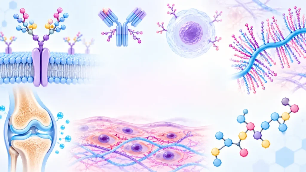
- Glycoproteins and proteoglycans are important carbohydrate-containing molecules found in almost all cells and tissues.
- They are made up of proteins and carbohydrate chains, but the amount, type, and arrangement of carbohydrates are different in the two groups.
- They are important for cell structure, cell recognition, signaling, immunity, extracellular matrix formation, and lubrication.
-
- Glycoproteins contain a protein molecule with relatively short and branched carbohydrate chains attached to it.
- Proteoglycans contain a core protein with long, repeating carbohydrate chains called glycosaminoglycans (GAGs).
- In glycoproteins, carbohydrates usually make up a smaller proportion of the molecule.
- In proteoglycans, carbohydrates can make up a large proportion of the molecule.
- Glycoproteins are commonly present on cell membranes, in blood, and in secretions.
- Proteoglycans are major components of the extracellular matrix (ECM).
- Both molecules participate in cell-cell recognition and communication.
Glycoproteins
A glycoprotein is a protein to which one or more carbohydrate chains are covalently attached. The carbohydrate portion is usually composed of oligosaccharides, which are relatively short carbohydrate chains.
Basic Structure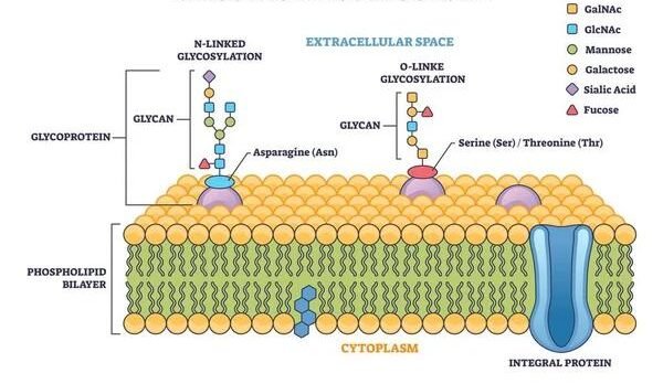
Protein + Oligosaccharide → Glycoprotein
The carbohydrate chains are attached to specific amino acid residues of the protein.
Structure of Glycoproteins
Glycoproteins consist of:
- Protein backbone
- Carbohydrate chains
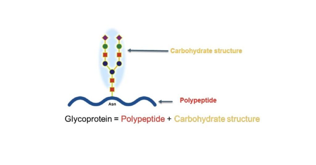
The carbohydrate portion may contain:
- Glucose
- Galactose
- Mannose
- Fucose
- N-acetylglucosamine (GlcNAc)
- N-acetylgalactosamine (GalNAc)
- Sialic acid
The exact carbohydrate composition differs from one glycoprotein to another.
Types of Glycoprotein Linkages
Carbohydrates can be attached to proteins mainly through N-linked or O-linked glycosylation.
A. N-Linked Glycosylation
- The carbohydrate chain is attached to the nitrogen atom of the amide group of asparagine.
- The basic recognition sequence is:
Asn–X–Ser/Thr
where X can be most amino acids except proline.
- N-linked glycosylation begins in the endoplasmic reticulum.
- Further processing commonly occurs in the Golgi apparatus.
Important
N-linked → Asparagine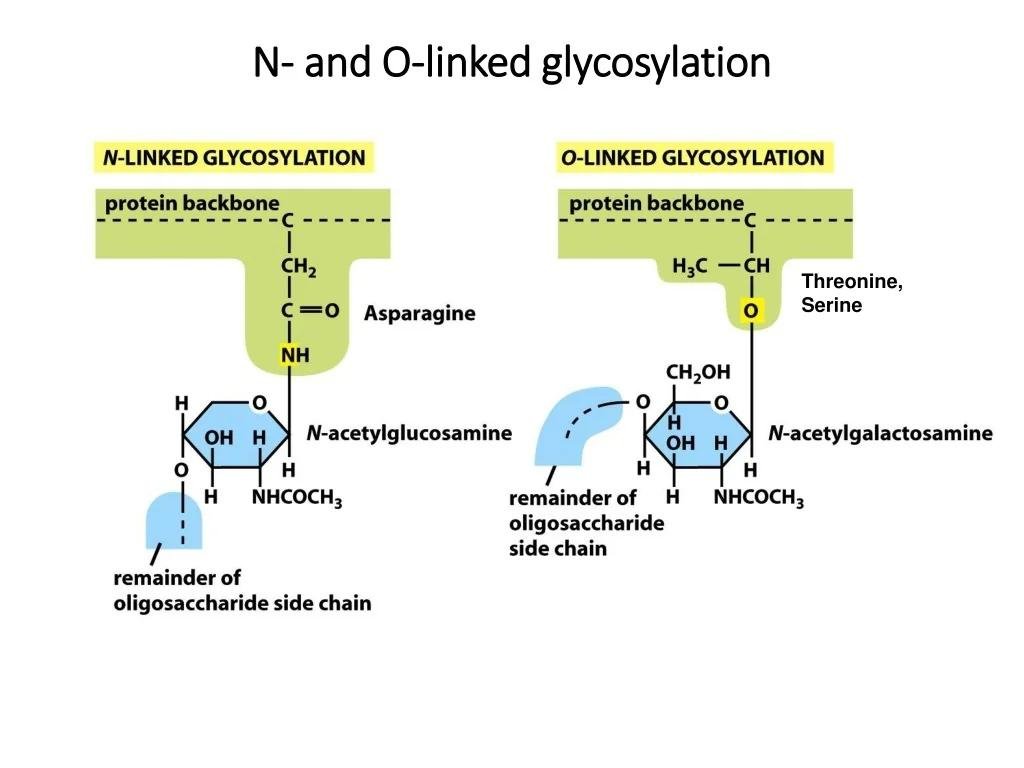
B. O-Linked Glycosylation
- The carbohydrate is attached to the oxygen atom of the hydroxyl group of:
- Serine
- Threonine
- O-linked glycosylation occurs mainly in the Golgi apparatus.
Important
O-linked → Serine or Threonine
Functions of Glycoproteins
Glycoproteins perform many important functions in the body. The carbohydrate part helps proteins in recognition, stability, communication, and protection.
- Cell Recognition – Help cells recognize and identify each other.
- Cell Adhesion – Help cells attach to other cells and to the extracellular matrix.
- Receptors – Many cell-surface receptors are glycoproteins that receive signals from hormones, neurotransmitters, and growth factors.
- Immune Function – Immunoglobulins (antibodies) and many immune-related proteins are glycoproteins that help protect the body from infections.
- Hormonal Functions – Several hormones, such as TSH, FSH, LH, and hCG, are glycoproteins.
- Transport – Some glycoproteins transport substances in the blood. For example, transferrin transports iron.
- Protection – Glycoproteins present in mucus help protect epithelial surfaces from microorganisms and physical damage.
- Blood Group Determination – Carbohydrate structures on glycoproteins and glycolipids contribute to ABO blood group antigens.
- Protein Stability – Glycosylation can improve the stability, folding, and lifespan of proteins.
- Cell Signaling – Glycoproteins participate in communication between cells and regulate processes such as growth and differentiation.
Examples of Glycoproteins
| Glycoprotein | Main Function |
|---|---|
| Immunoglobulins (Antibodies) | Immune defense against infections |
| TSH | Stimulates the thyroid gland |
| FSH | Regulates ovarian and testicular functions |
| LH | Regulates ovulation and reproductive functions |
| hCG | Maintains pregnancy in early stages |
| Erythropoietin (EPO) | Stimulates red blood cell production |
| Transferrin | Transports iron in the blood |
| Thyroxine-binding globulin (TBG) | Transports thyroid hormones |
| α₁-Acid glycoprotein | Transport and acute-phase response |
| Mucins | Protect and lubricate mucosal surfaces |
| Fibronectin | Cell adhesion and extracellular matrix organization |
| Laminin | Helps cells attach to the basement membrane |
| Many cell-surface receptors | Receive and transmit cellular signals |
Proteoglycans
Proteoglycans are molecules consisting of a core protein attached to one or more long glycosaminoglycan (GAG) chains.
Basic Structure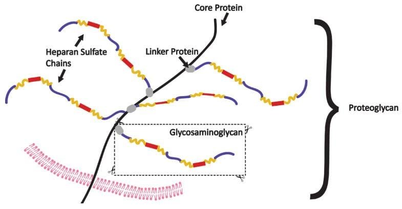
Core protein + GAG chains → Proteoglycan
The GAG chains are long, unbranched polysaccharides made up of repeating disaccharide units.
Structure of Proteoglycans
A typical proteoglycan contains:
- Core protein
- Several GAG chains
- Linkage region connecting GAGs to the protein
The GAG chains are usually highly negatively charged because they contain:
- Sulfate groups
- Carboxyl groups
This negative charge allows proteoglycans to attract water and positively charged ions.
Glycosaminoglycans
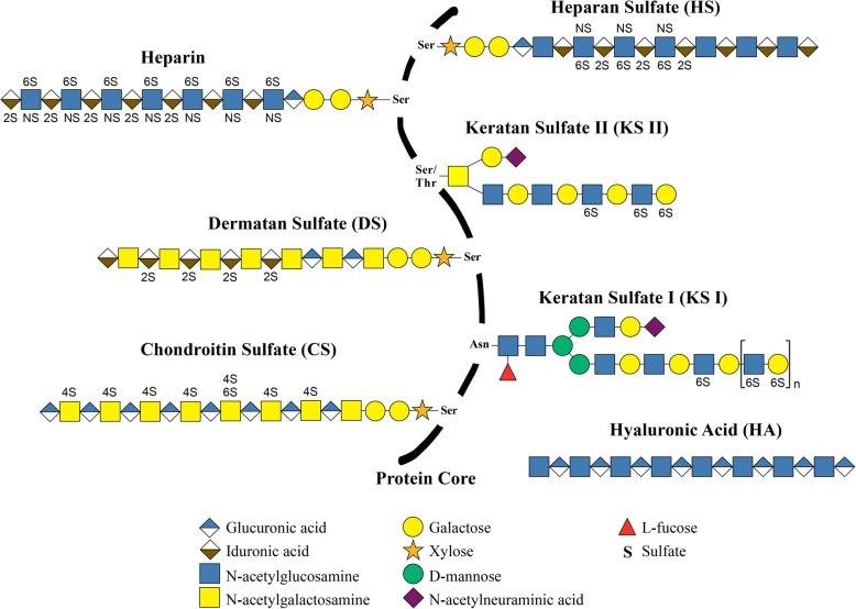
Glycosaminoglycans (GAGs) are long, unbranched carbohydrate chains made of repeating disaccharide units. They are important components of the extracellular matrix (ECM) and help provide structure, hydration, and lubrication to tissues.
Structure of GAGs
- GAGs are made of repeating two-sugar (disaccharide) units.
- Usually, one sugar is an amino sugar such as glucosamine or galactosamine.
- The other is usually a uronic acid or galactose.
- Most GAGs are negatively charged because they contain sulfate groups and/or carboxyl groups.
- Their negative charge allows them to attract water and positively charged ions.
- This property helps tissues maintain hydration and resistance to compression.
Major Types of GAGs
| GAG | Important Location/Function |
|---|---|
| Hyaluronic acid | Connective tissue, synovial fluid; lubrication |
| Chondroitin sulfate | Cartilage, bone, tendons; provides strength |
| Dermatan sulfate | Skin, blood vessels; tissue structure |
| Heparan sulfate | Cell surfaces and basement membranes; cell signaling |
| Heparin | Mast cells; strong anticoagulant activity |
| Keratan sulfate | Cornea, cartilage; structural support |
Functions of GAGs
- Maintain tissue hydration by attracting water.
- Provide lubrication, especially in joints.
- Resist compression in cartilage and connective tissues.
- Support the extracellular matrix.
- Help in cell adhesion and migration.
- Participate in cell signaling.
- Help regulate blood coagulation, especially heparin.
- Support wound healing and tissue repair.
Functions of Proteoglycans
Proteoglycans are important components of the extracellular matrix (ECM). They help provide structure, hydration, lubrication, and support to tissues.
- Provide Structural Support – Help maintain the structure and organization of connective tissues.
- Maintain Tissue Hydration – Their negatively charged GAG chains attract and hold large amounts of water.
- Resist Compression – Proteoglycans help cartilage withstand pressure and compression during movement.
- Provide Lubrication – They help reduce friction between tissues, especially in joints.
- Support the Extracellular Matrix – Work with collagen, elastin, and other ECM components to maintain tissue strength.
- Cell Adhesion – Help cells attach to the extracellular matrix and surrounding structures.
- Cell Signaling – Bind growth factors and signaling molecules and help regulate cellular activities.
- Cell Migration – Help regulate the movement of cells during development and tissue repair.
- Regulate Tissue Growth and Development – Participate in processes such as cell growth, differentiation, and tissue formation.
- Role in Blood Coagulation – Heparin, a highly sulfated GAG, has strong anticoagulant activity.
- Wound Healing and Tissue Repair – Help organize the extracellular matrix during tissue repair.
Proteoglycan Aggregates
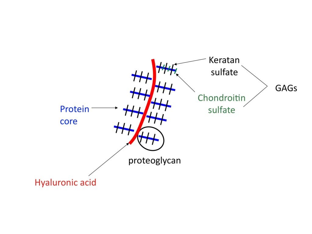
Proteoglycan aggregates are large complexes found mainly in the extracellular matrix of cartilage. They are formed when many proteoglycan molecules, especially aggrecan, attach to a long molecule of hyaluronic acid (hyaluronan).
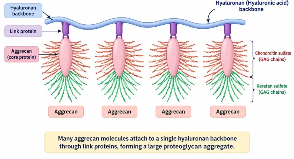 A proteoglycan aggregate mainly contains:
- Hyaluronic acid (Hyaluronan) – Acts as the long central backbone.
- Link proteins – Help attach proteoglycan molecules securely to hyaluronan.
- Aggrecan molecules – Large proteoglycans attached along the hyaluronan backbone.
- GAG chains – Chondroitin sulfate and keratan sulfate chains extend from the aggrecan core protein.
- Water – The negatively charged GAG chains attract and retain large amounts of water.
Functions
- Retain water in the cartilage matrix.
- Resist compression during movement and weight-bearing.
- Provide elasticity and cushioning to cartilage.
- Help maintain the structure of the extracellular matrix.
- Reduce friction and support smooth joint movement.
Glycoproteins vs Proteoglycans
| Feature | Glycoproteins | Proteoglycans |
|---|---|---|
| Basic structure | Protein + short carbohydrate chains | Core protein + GAG chains |
| Carbohydrate | Usually smaller proportion | Usually very large proportion |
| Carbohydrate type | Mainly oligosaccharides | Glycosaminoglycans |
| GAG chains | Absent | Present |
| Carbohydrate chains | Usually short and branched | Long and generally unbranched |
| Main location | Cell membrane, blood, secretions | Extracellular matrix |
| Main functions | Recognition, receptors, immunity, transport | Structure, hydration, compression resistance |
| Examples | Immunoglobulins, transferrin, TSH | Aggrecan, syndecans, perlecan |
Biosynthesis of Glycoproteins
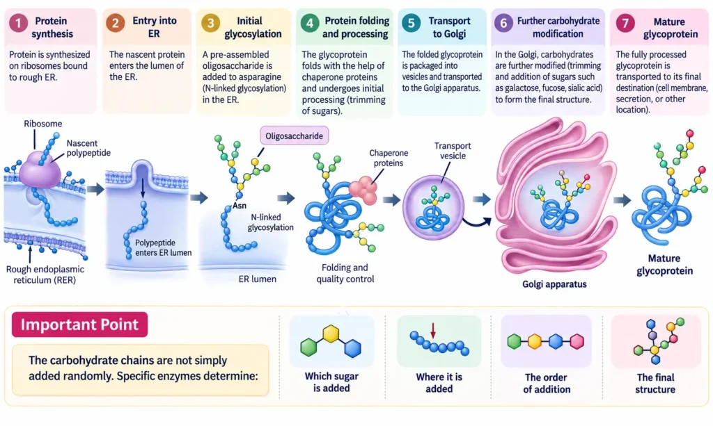
Glycoprotein synthesis mainly involves the endoplasmic reticulum and Golgi apparatus.
Steps
- Protein Synthesis
- The protein is synthesized by ribosomes attached to the rough ER.
- The newly formed polypeptide enters the ER lumen.
- Initial Glycosylation
- In the ER, carbohydrate chains are attached to specific amino acid residues of the protein.
- N-linked glycosylation occurs mainly on the amino acid asparagine (Asn).
- A preformed oligosaccharide is transferred to the protein.
- Protein Folding and Processing
- The glycoprotein folds into its proper three-dimensional structure.
- Chaperone proteins help proper folding.
- Some sugars may be removed or modified during this process.
- Transport to Golgi
- The partially processed glycoprotein is packed into transport vesicles.
- These vesicles carry it from the ER to the Golgi apparatus.
- Further Carbohydrate Modification
- In the Golgi, carbohydrate chains undergo further modification.
- Specific sugars may be added or removed.
- Sugars such as galactose, fucose, and sialic acid may be incorporated.
- O-Linked Glycosylation
- In many glycoproteins, carbohydrates can also be attached to the oxygen of serine or threonine.
- This type of glycosylation mainly occurs in the Golgi apparatus.
- Formation of Mature Glycoprotein
- After processing is complete, the mature glycoprotein is packed into vesicles.
- It is transported to its final location, such as the cell membrane, lysosome, or extracellular space.
Biosynthesis of Proteoglycans
 Steps
Steps
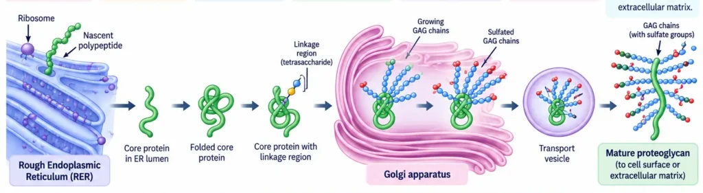
- Core Protein Synthesis
- The core protein is synthesized by ribosomes on the rough ER.
- The newly formed protein enters the ER.
- Protein Folding and Processing
- The core protein folds into its proper shape.
- Initial modifications may occur before it moves to the Golgi.
- Formation of the Linkage Region
- A specific carbohydrate linkage region is attached to certain serine residues of the core protein.
- This linkage serves as the starting point for GAG chain formation.
- GAG Chain Formation
- In the Golgi apparatus, sugar molecules are added one by one.
- This produces long, repeating GAG chains such as chondroitin sulfate, dermatan sulfate, and heparan sulfate.
- Sulfation and Modification
- Sulfate groups are added to the GAG chains.
- These modifications give GAGs their strong negative charge and help determine their biological functions.
- Final Processing
- The proteoglycan undergoes final processing and quality control.
- Its structure and carbohydrate composition are determined by specific enzymes.
- Transport and Secretion
- The mature proteoglycan is packed into vesicles.
- It is transported to the cell surface or extracellular matrix.
Most proteoglycan processing occurs through the ER and Golgi pathway.
Role in Extracellular Matrix
- Structural Support – Glycoproteins and proteoglycans help maintain the structure and organization of tissues.
- Cell Adhesion – Glycoproteins such as fibronectin and laminin help cells attach to the ECM.
- Hydration – Proteoglycans attract and retain water through their negatively charged GAG chains.
- Resistance to Compression – Proteoglycans, especially aggrecan, help cartilage resist pressure and compression.
- Organization of Collagen – ECM glycoproteins help organize collagen fibers and maintain tissue strength.
- Cell Migration – ECM components provide a surface that helps cells move during development and wound healing.
- Cell Signaling – Proteoglycans can bind growth factors and other signaling molecules, helping regulate cell behavior.
- Tissue Repair – ECM components participate in wound healing and tissue regeneration.
- Basement Membrane Formation – Laminin and other glycoproteins are important components of basement membranes.
- Lubrication and Cushioning – Proteoglycans and hyaluronic acid help provide lubrication and cushioning, particularly in joints and cartilage.
Clinical Significance
Glycoproteins and proteoglycans are important for cell function, tissue structure, immunity, signaling, and disease processes. Abnormal synthesis or breakdown of these molecules can lead to various disorders.
- Congenital Disorders of Glycosylation (CDG) – Defects in glycoprotein glycosylation can affect many organs and may cause developmental, neurological, and metabolic problems.
- Lysosomal Storage Disorders – Defects in the breakdown of GAGs can cause their accumulation in tissues, as seen in mucopolysaccharidoses.
- Osteoarthritis – Loss and breakdown of proteoglycans such as aggrecan reduce the water-holding capacity and compressive strength of cartilage.
- Cancer – Changes in cell-surface glycoproteins and proteoglycans can affect cell adhesion, migration, growth, and tumor progression.
- Inflammation – Proteoglycans can interact with inflammatory mediators and influence inflammatory responses.
- Immune Disorders – Abnormal glycoproteins can affect antibody function, immune-cell recognition, and other immune processes.
- Blood Coagulation – Heparin, a highly sulfated GAG, has important anticoagulant activity and is widely used clinically to prevent blood clot formation.
- Kidney Diseases – Changes in basement-membrane glycoproteins and proteoglycans can affect the glomerular filtration barrier.
- Inherited Metabolic Disorders – Enzyme defects affecting GAG synthesis or degradation can lead to abnormal accumulation of GAGs in tissues.
- Wound Healing – Alterations in ECM glycoproteins and proteoglycans can influence cell migration, tissue repair, and regeneration.