Introduction
- Cancer is a group of diseases in which abnormal cells grow and divide uncontrollably.
- Normally, cells grow, divide, and die in a controlled manner.
- In cancer, changes in genes and cellular signaling disturb normal cell regulation.
- These changes allow abnormal cells to survive and multiply excessively.
- Cancer can develop in almost any tissue or organ of the body.
- Cancer cells may invade nearby tissues and can spread to other parts of the body. This spread is called metastasis.
- Important changes in cancer involve oncogenes, tumor suppressor genes, DNA repair genes, and cell-cycle regulators.
- Cancer cells can also avoid apoptosis (programmed cell death) and continue to survive.
- Some cancer cells stimulate the formation of new blood vessels, a process called angiogenesis, to support their growth.
- Cancer biology helps us understand how normal cells become cancerous and how cancer develops, progresses, and spreads.
Incidence of Cancer
- Cancer is one of the major causes of illness and death worldwide.
- Cancer can occur in people of all ages, although the risk generally increases with age.
- The incidence of cancer varies between countries, regions, age groups, and sexes.
- Common cancers worldwide include breast, lung, colorectal, prostate, and stomach cancers.
- In males, lung, prostate, and colorectal cancers are among the commonly diagnosed cancers.
- In females, breast, lung, and colorectal cancers are among the commonly diagnosed cancers.
- In India, commonly reported cancers include breast, cervical, oral, lung, and colorectal cancers, with variation across different regions.
- Cancer incidence is influenced by factors such as age, genetics, tobacco use, alcohol consumption, diet, infections, obesity, environmental exposures, and lifestyle.
- Improvements in cancer screening and diagnostic methods can increase the number of cancers detected.
Etiology of Cancer
Cancer does not have a single cause. It usually develops due to a combination of genetic changes, environmental exposures, infections, and lifestyle factors.
1. Genetic Factors
- Inherited mutations can increase the risk of certain cancers.
- Mutations may also develop during a person’s lifetime.
- Changes in oncogenes, tumor suppressor genes, and DNA repair genes can contribute to cancer.
2. Tobacco Use
- Smoking and other forms of tobacco are major causes of cancer.
- Tobacco is strongly associated with cancers of the lung, oral cavity, throat, and urinary bladder.
3. Alcohol Consumption
- Excessive alcohol consumption can increase the risk of several cancers.
- The risk may be higher when alcohol use is combined with tobacco.
4. Infections
Certain infections can contribute to cancer development, including:
- Human papillomavirus (HPV) → cervical and other cancers
- Hepatitis B and C viruses → liver cancer
- Helicobacter pylori → stomach cancer
- Epstein–Barr virus (EBV) → some lymphomas and other cancers
5. Radiation
- Ionizing radiation can damage cellular DNA.
- Excessive ultraviolet (UV) radiation increases the risk of skin cancer.
6. Chemical Carcinogens
Exposure to certain chemicals can cause DNA damage and increase cancer risk. Examples include:
- Asbestos
- Benzene
- Certain industrial chemicals
- Some occupational carcinogens
7. Diet and Lifestyle
- Unhealthy dietary patterns may increase the risk of certain cancers.
- Obesity, physical inactivity, and low consumption of fruits and vegetables can also contribute to cancer risk.
8. Hormonal Factors
- Prolonged exposure to certain hormones can influence the development of some cancers.
- Hormonal factors are particularly important in some breast and reproductive system cancers.
9. Chronic Inflammation
- Long-term inflammation can cause repeated cellular injury and DNA damage.
- Chronic inflammatory conditions may increase the risk of certain cancers.
10. Age
- Cancer risk generally increases with age.
- This is partly because genetic and cellular damage can accumulate over many years.
11. Environmental and Occupational Factors
- Air pollution and certain environmental exposures may contribute to cancer development.
- Occupational exposure to radiation, chemicals, dust, and other carcinogens can also increase risk.
12. Random Cellular Changes
- Some DNA mutations occur naturally during normal cell division.
- In some cases, these random changes may contribute to cancer development even when no specific external cause is identified.
Molecular Basis of Cancer
Cancer develops when normal cells acquire genetic and molecular abnormalities that disturb the normal control of cell growth, division, differentiation, and death. These changes occur gradually and involve multiple genes and signaling pathways.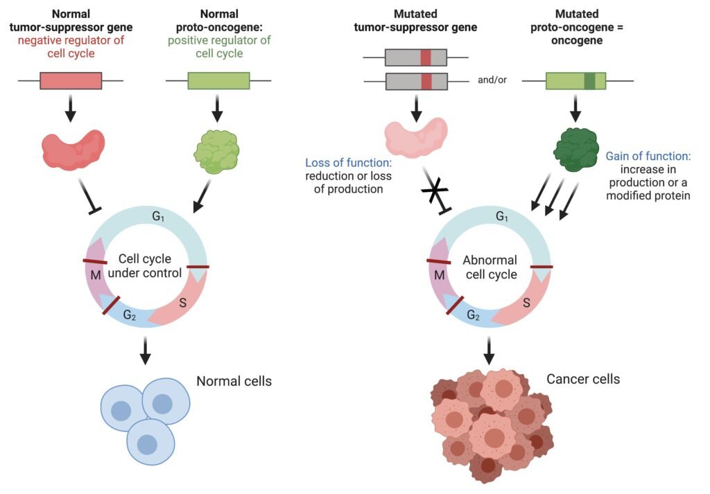
1. Genetic Alterations
Genetic alterations are the fundamental events involved in cancer development.
- Mutations may develop due to:
- DNA replication errors
- Tobacco and chemical exposure
- Radiation
- Certain infections
- Oxidative stress
- Inherited genetic defects
- These alterations can affect genes controlling:
- Cell proliferation
- Cell-cycle regulation
- DNA repair
- Apoptosis
- Cell differentiation
- Cell adhesion and migration
- Accumulation of these changes gradually converts a normal cell into a malignant cell.
2. Activation of Oncogenes
Proto-oncogenes are normal genes that promote cell growth and division when required.
- Genetic alterations can convert proto-oncogenes into oncogenes.
- Oncogenes may become activated through:
- Point mutations
- Gene amplification
- Chromosomal translocation
- Increased gene expression
- Activated oncogenes continuously stimulate cell proliferation.
- Important oncogenes include:
- RAS
- MYC
- HER2
- BCR-ABL
Example: RAS
- RAS normally transmits growth signals from cell-surface receptors.
- Mutated RAS may remain continuously active.
- Continuous RAS signaling activates pathways such as the MAPK pathway.
- This promotes:
- Cell proliferation
- Cell survival
- Tumor growth
3. Loss of Tumor Suppressor Genes
Tumor suppressor genes act as natural brakes on cell growth.
- They normally:
- Prevent uncontrolled cell division
- Promote cell-cycle arrest
- Facilitate DNA repair
- Promote apoptosis when damage is severe
- Important tumor suppressor genes include:
- TP53
- RB1
- APC
- PTEN
- Loss or inactivation of these genes removes important controls over cell proliferation.
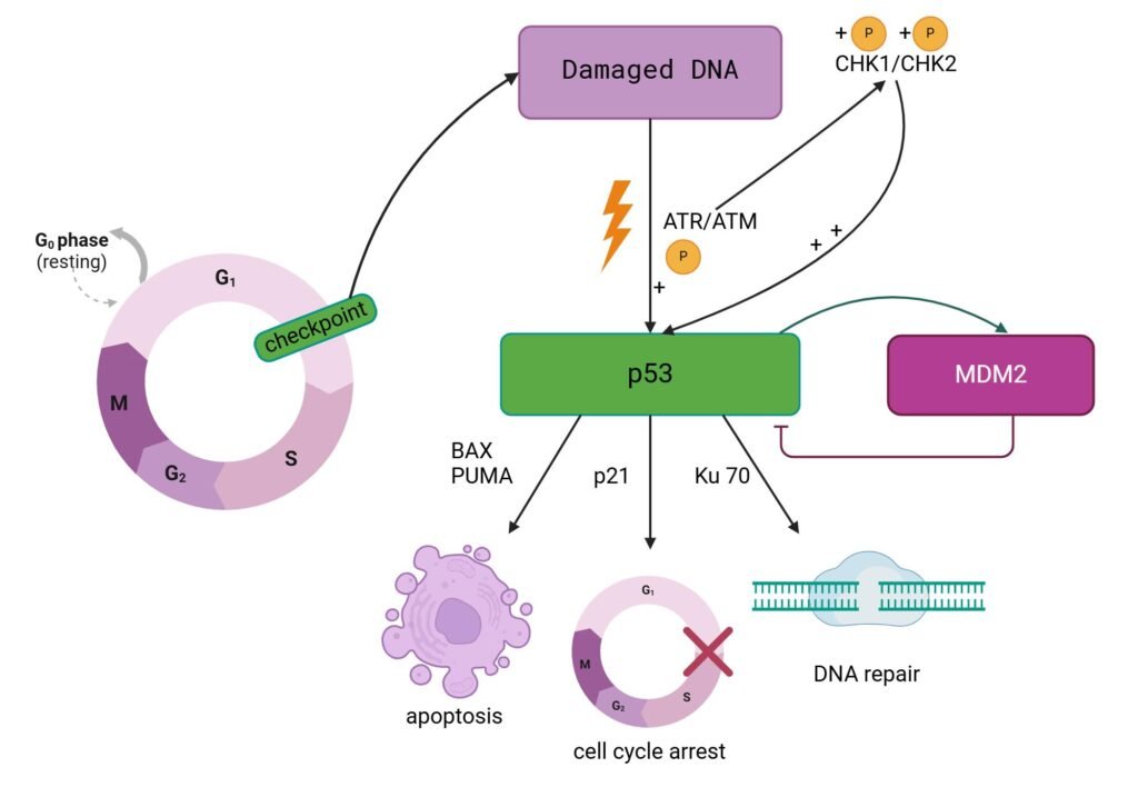
Role of TP53
- TP53 encodes the p53 protein.
- p53 responds to cellular stress and DNA damage.
- It can:
- Stop the cell cycle
- Allow DNA repair
- Promote apoptosis if damage is severe
- Loss of p53 function allows damaged cells to survive and continue dividing.
- This increases the accumulation of additional genetic abnormalities.
4. Abnormal Cell-Cycle Regulation
The cell cycle is normally controlled by several checkpoints.
- Important regulators include:
- Cyclins
- Cyclin-dependent kinases (CDKs)
- RB protein
- p53
- The G1/S checkpoint is particularly important.
- RB normally regulates the activity of E2F transcription factors.
- Loss of RB control allows E2F to remain active.
- This promotes:
- DNA synthesis
- S-phase entry
- Repeated cell division
- As a result, cancer cells may continue dividing despite inappropriate conditions.
5. Abnormal Growth Factor Signaling
Normal cells require growth signals for proliferation.
- Growth factors bind to specific receptors on the cell surface.
- These receptors activate intracellular signaling pathways.
- Important pathways include:
- RAS–RAF–MEK–ERK (MAPK)
- PI3K–AKT–mTOR
- JAK–STAT
- Wnt/β-catenin
- Genetic abnormalities can cause these pathways to remain continuously active.
- This results in:
- Increased cell proliferation
- Increased cell survival
- Increased cellular growth
6. PI3K–AKT–mTOR Pathway
The PI3K–AKT–mTOR pathway regulates cell growth, metabolism, protein synthesis, and survival.
- Growth factor stimulation can activate PI3K.
- PI3K activates AKT.
- AKT promotes mTOR activity.
- mTOR stimulates:
- Protein synthesis
- Cell growth
- Cellular metabolism
- Cell survival
- Excessive activation of this pathway can promote cancer development.
- PTEN normally acts as an important negative regulator of this pathway.
- Loss of PTEN can therefore enhance cancer-promoting signaling.
7. Evasion of Apoptosis
Apoptosis is programmed cell death that removes damaged or unnecessary cells.
- Normal cells with severe DNA damage may undergo apoptosis.
- Cancer cells can escape apoptosis through:
- Loss of p53 activity
- Increased anti-apoptotic proteins such as BCL-2
- Reduced pro-apoptotic signaling
- Alteration of caspase pathways
- As a result:
- Damaged cells survive
- Abnormal cells accumulate
- Tumor growth continues
8. Defective DNA Repair
DNA repair mechanisms protect cells from the accumulation of genetic damage.
- Important DNA repair genes include:
- BRCA1
- BRCA2
- MLH1
- MSH2
- MSH6
- PMS2
- Defects in DNA repair lead to:
- Accumulation of mutations
- Genomic instability
- Increased risk of malignant transformation
- DNA repair defects may also increase mutations in oncogenes and tumor suppressor genes.
9. Epigenetic Alterations
Epigenetic changes affect gene expression without changing the DNA sequence.
Important mechanisms include:
- DNA methylation
- Histone modification
- Chromatin remodeling
- Non-coding RNA regulation
Important effects
- Increased methylation of a tumor suppressor gene promoter can reduce gene expression.
- Reduced expression of tumor suppressor proteins can promote uncontrolled growth.
- Abnormal epigenetic changes can also alter differentiation and cellular metabolism.
10. Telomerase Activation
Telomeres protect the ends of chromosomes and normally become shorter with repeated cell division.
- Progressive telomere shortening eventually leads to cellular senescence or death.
- Many cancer cells increase telomerase activity.
- Telomerase helps maintain telomere length.
- This allows cancer cells to:
- Continue dividing
- Avoid replicative senescence
- Achieve prolonged or potentially unlimited replicative capacity
11. Tumor Angiogenesis
Growing tumors require an adequate supply of oxygen and nutrients.
- Rapidly growing tumors may develop areas of low oxygen.
- Hypoxia can stabilize HIF-1α.
- HIF-1α promotes the expression of angiogenic factors such as VEGF.
- VEGF stimulates new blood-vessel formation.
- Tumor angiogenesis provides:
- Oxygen
- Nutrients
- Growth support
- Access to the circulation
- It can also facilitate metastatic spread.
12. Invasion and Metastasis
Malignant cells can invade surrounding tissues and spread to distant organs.
Important changes include:
- Loss of normal cell-to-cell adhesion
- Reduced E-cadherin activity
- Increased cellular migration
- Degradation of extracellular matrix
- Entry into blood or lymphatic vessels
- Colonization of distant organs
Cancer cells can produce or stimulate enzymes such as matrix metalloproteinases (MMPs), which help break down extracellular matrix components.
13. Epithelial–Mesenchymal Transition
Epithelial–mesenchymal transition (EMT) is associated with increased migration and invasion of cancer cells.
During EMT:
- Epithelial characteristics are reduced.
- Cell-to-cell adhesion decreases.
- Cellular mobility increases.
- Invasive properties increase.
- Metastatic potential may increase.
Important regulatory factors include:
- SNAIL
- SLUG
- TWIST
- ZEB
14. Immune Evasion
The immune system can recognize and eliminate abnormal cells. Cancer cells may develop mechanisms to escape immune destruction.
- Some cancer cells express PD-L1.
- PD-L1 interacts with PD-1 receptors on T cells.
- This interaction reduces T-cell activity.
- Consequently, cancer cells may survive despite immune surveillance.
This mechanism has led to the development of immune checkpoint inhibitors used in the treatment of several cancers.
15. Role of Chronic Inflammation
Chronic inflammation can contribute to cancer development.
Persistent inflammation may produce:
- Reactive oxygen species
- Reactive nitrogen species
- Cytokines
- Growth factors
These factors can:
- Damage DNA
- Promote cell proliferation
- Alter tissue structure
- Support survival of abnormal cells
Important inflammatory signaling pathways include NF-κB and STAT3.
16. Tumor Microenvironment
A tumor contains not only cancer cells but also several surrounding cell types and extracellular components.
The tumor microenvironment may include:
- Fibroblasts
- Endothelial cells
- Immune cells
- Extracellular matrix
- Cytokines
- Growth factors
These components can support:
- Tumor growth
- Angiogenesis
- Invasion
- Metastasis
- Immune evasion
- Treatment resistance
Tumor Markers
- Tumor markers are substances produced by cancer cells or by normal cells in response to cancer.
- They may be found in blood, urine, body fluids, or tumor tissues.
- Tumor markers are useful in cancer diagnosis, monitoring treatment, detecting recurrence, and assessing prognosis.
- However, most tumor markers are not specific enough to diagnose cancer by themselves and should be interpreted along with clinical findings, imaging, and histopathology.
Types of Tumor Markers
- Tumor markers can broadly be classified into:
A. Biochemical Tumor Markers
- These are substances that can be measured in body fluids, particularly blood.
Examples include:
- AFP (Alpha-fetoprotein)
- CEA (Carcinoembryonic antigen)
- PSA (Prostate-specific antigen)
- CA-125
- CA 19-9
- CA 15-3
- β-hCG
B. Molecular and Genetic Tumor Markers
- These are specific genes, mutations, proteins, or molecular alterations associated with cancer.
Examples include:
- HER2
- EGFR
- KRAS
- BRAF
- BRCA1/BRCA2
- ALK
- PD-L1
These markers are particularly important for targeted therapy and personalized cancer treatment.
Important Tumor Markers
| Tumor Marker | Commonly Associated Cancer | Major Clinical Use |
|---|---|---|
| AFP | Hepatocellular carcinoma, germ-cell tumors | Diagnosis support and monitoring |
| CEA | Colorectal and other cancers | Monitoring and recurrence |
| PSA | Prostate cancer | Screening support, monitoring |
| CA-125 | Ovarian cancer | Monitoring and recurrence |
| CA 19-9 | Pancreatic and biliary cancers | Monitoring |
| CA 15-3 | Breast cancer | Monitoring |
| β-hCG | Gestational trophoblastic disease, germ-cell tumors | Diagnosis and monitoring |
| Calcitonin | Medullary thyroid carcinoma | Diagnosis and monitoring |
| Thyroglobulin | Differentiated thyroid cancer | Follow-up after treatment |
| Chromogranin A | Neuroendocrine tumors | Assessment and monitoring |
Alpha-Fetoprotein (AFP)
- AFP is a major fetal plasma protein produced mainly by the fetal liver.
Clinical significance
- Increased AFP is commonly associated with:
- Hepatocellular carcinoma
- Certain germ-cell tumors
- AFP may also increase in some non-malignant liver diseases, including hepatitis and cirrhosis.
- Therefore, an elevated AFP level alone does not confirm cancer.
Carcinoembryonic Antigen (CEA)
- CEA is a glycoprotein associated particularly with embryonic and gastrointestinal tissues.
Clinical significance
- Commonly associated with:
- Colorectal cancer
- Pancreatic cancer
- Gastric cancer
- Some other malignancies
- CEA is mainly useful for:
- Monitoring treatment
- Detecting possible recurrence
- Assessing disease progression
CEA may also be elevated in some benign conditions and in smokers.
Prostate-Specific Antigen (PSA)
- PSA is a serine protease produced mainly by prostate epithelial cells.
Clinical significance
- PSA can be increased in:
- Prostate cancer
- Benign prostatic hyperplasia
- Prostatitis
- PSA is used in conjunction with other clinical assessments for prostate cancer detection and risk assessment.
- Serial PSA measurements can also help monitor patients after treatment.
CA-125
- CA-125 is a high-molecular-weight glycoprotein that is particularly associated with epithelial ovarian cancer.
Clinical significance
- It is mainly used for:
- Monitoring ovarian cancer
- Assessing response to treatment
- Detecting possible recurrence
- CA-125 can also increase in several benign gynecological and inflammatory conditions.
Therefore, it is not a stand-alone diagnostic test for ovarian cancer.
CA 19-9
- CA 19-9 is a carbohydrate-associated antigen.
Clinical significance
It may be elevated in:
- Pancreatic cancer
- Biliary tract cancers
- Some gastrointestinal cancers
It is mainly useful for monitoring disease and treatment response rather than establishing a diagnosis by itself.
CA 15-3
- CA 15-3 is associated particularly with breast cancer.
Clinical significance
- It may be used to monitor patients with advanced breast cancer.
- Changes in its concentration may provide information about:
- Treatment response
- Disease progression
- Recurrence
It is not recommended as a stand-alone screening test for breast cancer.
β-hCG
- β-human chorionic gonadotropin (β-hCG) is a hormone normally produced during pregnancy.
Tumor-related significance
Elevated β-hCG can occur in:
- Gestational trophoblastic disease
- Choriocarcinoma
- Some germ-cell tumors
It can be particularly useful for diagnosis and monitoring treatment response.
Molecular Tumor Markers
- Modern cancer diagnosis increasingly uses molecular markers to identify specific alterations within tumor cells.
Important examples
HER2
- Commonly assessed in breast and certain other cancers.
- HER2 overexpression or amplification can influence treatment selection.
EGFR
- Mutations in EGFR are important in some non-small-cell lung cancers.
- Certain EGFR alterations can help identify patients who may benefit from targeted therapy.
KRAS
- KRAS mutations occur in several cancers, including colorectal and lung cancers.
- KRAS status can influence the use of certain targeted therapies.
BRAF
- BRAF mutations, particularly BRAF V600E, occur in melanoma and several other cancers.
- BRAF status can guide targeted treatment in appropriate patients.
BRCA1 and BRCA2
- Alterations in these genes impair DNA repair.
- They are relevant to hereditary breast and ovarian cancer and also have therapeutic implications in several cancers.
Uses of Tumor Markers
Tumor markers have several clinical applications.
1. Screening
- Some markers may have a role in selected populations, but most tumor markers are not suitable for general population screening because of limited sensitivity or specificity.
2. Supporting Diagnosis
- Tumor markers can provide additional evidence when cancer is suspected.
- However, histopathological examination and other investigations remain essential for definitive diagnosis in most cancers.
3. Monitoring Treatment
- Changes in tumor-marker concentrations can help assess whether a tumor is responding to treatment.
4. Detecting Recurrence
- After successful treatment, a rising marker level may suggest recurrent disease and may prompt further evaluation.
5. Prognostic Assessment
- Some tumor markers provide information about the likely behavior or progression of a cancer.
6. Selecting Targeted Therapy
- Molecular markers can identify specific abnormalities that may make a tumor suitable for a particular targeted treatment.
Limitations of Tumor Markers
Tumor markers have important limitations.
- Not all cancers produce detectable tumor markers.
- A marker may be elevated in non-cancerous conditions.
- The same marker may be increased in different types of cancer.
- A normal marker level does not always exclude cancer.
- Elevated levels do not necessarily prove the presence of cancer.
- Results should be interpreted together with:
- Clinical findings
- Imaging
- Histopathology
- Other laboratory investigations
Characteristics of Growing Tumor Cells
Tumor cells differ from normal cells in their growth pattern, metabolism, differentiation, survival, and interaction with surrounding tissues. As genetic and molecular abnormalities accumulate, tumor cells acquire characteristics that allow them to survive and multiply continuously.
1. Uncontrolled Cell Proliferation
- Tumor cells divide more frequently than normal cells.
- They may continue to proliferate even when growth signals are absent.
- Normal mechanisms that limit cell division become ineffective.
- This results in progressive accumulation of abnormal cells.
2. Reduced Growth Control
- Normal cells respond to signals that promote or inhibit growth.
- Tumor cells may become less responsive to growth-inhibitory signals.
- Alterations in oncogenes and tumor suppressor genes contribute to this loss of control.
- As a result, cells continue to grow despite unfavorable conditions.
3. Resistance to Apoptosis
- Normal damaged cells can undergo apoptosis.
- Tumor cells may develop mechanisms to avoid programmed cell death.
- This can occur through:
- Loss of p53 activity
- Increased anti-apoptotic proteins such as BCL-2
- Altered caspase signaling
- Damaged cells therefore survive and continue to proliferate.
4. Replicative Immortality
- Normal cells have a limited number of cell divisions.
- Many tumor cells activate telomerase.
- Telomerase helps maintain telomere length.
- This allows tumor cells to continue dividing for prolonged periods.
- Therefore, cancer cells can acquire replicative immortality.
5. Loss of Normal Differentiation
- Normal cells usually develop specialized structures and functions.
- Tumor cells may become less differentiated.
- Some malignant tumors show marked anaplasia, characterized by:
- Cellular and nuclear pleomorphism
- Enlarged and irregular nuclei
- Abnormal nuclear-to-cytoplasmic ratio
- Abnormal mitotic figures
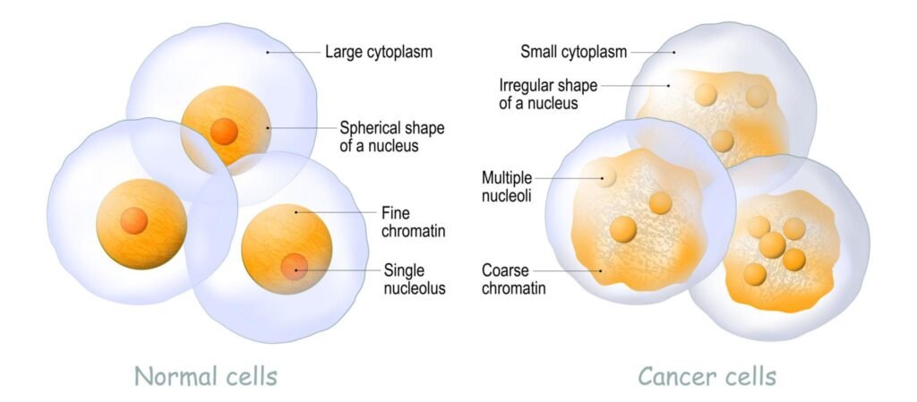
6. Abnormal Cellular Morphology
Growing tumor cells may show characteristic morphological changes.
Common findings include:
- Variation in cell size (pleomorphism)
- Variation in nuclear size and shape
- Hyperchromatic nuclei
- Prominent nucleoli
- Increased nuclear-to-cytoplasmic ratio
- Abnormal mitotic figures
These features are important in histopathological examination.
7. Altered Cellular Metabolism
Tumor cells frequently undergo metabolic reprogramming.
- Many cancer cells increase glucose uptake.
- They may preferentially use glycolysis even when oxygen is available, a phenomenon known as the Warburg effect.
- Increased glycolysis provides:
- ATP
- Biosynthetic intermediates
- Metabolic support for rapid cell proliferation
- This altered metabolism helps cancer cells meet their increased energy and biosynthetic requirements.
8. Increased Angiogenesis
- Rapidly growing tumors require increased oxygen and nutrients.
- Tumor cells can stimulate formation of new blood vessels.
- This process is called angiogenesis.
- VEGF is an important mediator.
- Increased vascular supply supports continued tumor growth.
9. Ability to Invade Surrounding Tissues
Malignant tumor cells can invade nearby tissues.
Important changes include:
- Reduced cell-to-cell adhesion
- Altered extracellular matrix interactions
- Increased production of matrix-degrading enzymes
- Increased cellular migration
This property distinguishes malignant tumors from most benign tumors.
10. Ability to Metastasize
Some tumor cells can spread from the primary tumor to distant organs.
The metastatic process involves:
- Local invasion
- Entry into blood or lymphatic vessels
- Survival in circulation
- Exit from the circulation
- Growth at a distant site
Metastasis is one of the most important characteristics of malignant tumors.
11. Immune Evasion
Tumor cells can develop mechanisms to avoid destruction by the immune system.
They may:
- Reduce recognition by immune cells
- Alter antigen expression
- Produce immunosuppressive signals
- Express immune checkpoint molecules such as PD-L1
This allows abnormal cells to survive despite immune surveillance.
12. Genomic Instability
- Tumor cells often accumulate numerous genetic abnormalities.
- Defects in DNA repair mechanisms contribute to this process.
- Genomic instability promotes additional mutations.
- These mutations can further enhance tumor growth, survival, invasion, and treatment resistance.
13. Clonal Evolution
Cancer cells within the same tumor are not always genetically identical.
- Genetic changes occur continuously during tumor growth.
- Cells with advantageous mutations may survive and multiply more successfully.
- Over time, these cell populations become dominant.
- This process is called clonal evolution.
It contributes to:
- Tumor progression
- Heterogeneity
- Metastasis
- Treatment resistance
14. Tumor Heterogeneity
A tumor may contain several populations of cells with different genetic and molecular characteristics.
This tumor heterogeneity can affect:
- Growth rate
- Metastatic potential
- Response to treatment
- Development of drug resistance
15. Interaction with the Tumor Microenvironment
Tumor cells interact continuously with surrounding cells and tissues.
The tumor microenvironment contains:
- Fibroblasts
- Immune cells
- Endothelial cells
- Extracellular matrix
- Cytokines
- Growth factors
These interactions can support tumor growth, angiogenesis, invasion, immune evasion, and progression.
Cancer Therapy
- Cancer therapy refers to the use of different medical approaches to control, eliminate, or slow the growth of cancer cells.
- Treatment is selected according to the type and stage of cancer, molecular characteristics of the tumor, location, overall clinical condition, and treatment goals.
Modern cancer treatment often combines more than one approach, such as surgery with chemotherapy, radiotherapy, targeted therapy, or immunotherapy.
1. Surgery
Surgery involves the physical removal of the tumor from the body.
- It is commonly used for localized solid tumors.
- The surgeon may remove:
- The primary tumor
- A margin of surrounding tissue
- Nearby lymph nodes when clinically indicated
- Surgery may be used with curative or palliative intent.
- It is often combined with other treatments.
Example
In some cancers, surgery is followed by chemotherapy or radiotherapy to reduce the risk of residual or recurrent disease.
2. Radiation Therapy
- Radiation therapy uses high-energy radiation to damage the DNA of cancer cells.
Mechanism
- Radiation produces direct and indirect DNA damage.
- This can cause:
- DNA strand breaks
- Chromosomal damage
- Loss of reproductive ability
- Cell death
- Cancer cells that cannot adequately repair the damage may undergo apoptosis or other forms of cell death.
Uses
Radiotherapy may be used:
- To treat localized cancer
- After surgery
- Before surgery in selected cancers
- Along with chemotherapy
- To relieve symptoms such as pain caused by advanced cancer
3. Chemotherapy
- Chemotherapy uses cytotoxic drugs that interfere with cancer-cell proliferation.
- Because many chemotherapy drugs act on rapidly dividing cells, they can also affect some normal rapidly dividing tissues.
Major mechanisms
Chemotherapeutic drugs may:
- Damage DNA
- Inhibit DNA synthesis
- Inhibit RNA or protein synthesis
- Interfere with mitosis
- Prevent cell division
Examples
- Alkylating agents
- Antimetabolites
- Anthracyclines
- Topoisomerase inhibitors
- Microtubule inhibitors
Chemotherapy may be administered before surgery (neoadjuvant therapy), after surgery (adjuvant therapy), or for advanced disease.
4. Hormonal Therapy
- Some cancers depend on hormones for growth.
- Hormonal therapy reduces hormonal stimulation or blocks hormone receptors.
Common examples
- Breast cancer: Estrogen or progesterone signaling may be targeted.
- Prostate cancer: Androgen signaling may be suppressed or blocked.
Mechanism
Hormonal therapy can:
- Reduce hormone production
- Block hormone receptors
- Prevent hormone-dependent signaling
This can slow the growth of hormone-sensitive cancer cells.
5. Targeted Therapy
- Targeted therapy is designed to act on specific molecular abnormalities or pathways involved in cancer.
- Unlike conventional chemotherapy, many targeted therapies are developed to interfere with particular molecules that are important for cancer-cell survival or proliferation.
Examples
HER2-targeted therapy
- Used in appropriate HER2-positive cancers.
- Targets abnormal HER2 signaling.
EGFR-targeted therapy
- Used in selected cancers with specific EGFR alterations.
BRAF-targeted therapy
- Used in cancers with certain BRAF mutations.
BCR-ABL-targeted therapy
- Important in chronic myeloid leukemia.
Advantages
- More selective molecular targeting
- Can produce significant responses in tumors with the appropriate molecular alteration
- Provides the basis for precision oncology
6. Immunotherapy
- Immunotherapy helps the immune system recognize or attack cancer cells.
- Immune Checkpoint Inhibitors
- Cancer cells can suppress T-cell activity through immune checkpoint pathways.
Important targets include:
- PD-1
- PD-L1
- CTLA-4
Checkpoint inhibitors block these inhibitory signals and can restore T-cell activity against cancer cells.
Other Immunotherapy Approaches
- Monoclonal antibodies
- Cancer vaccines
- Adoptive cell therapy
- CAR-T cell therapy
The effectiveness of immunotherapy varies according to cancer type and individual tumor characteristics.
7. Monoclonal Antibody Therapy
- Monoclonal antibodies are laboratory-produced antibodies designed to recognize specific molecules.
They can work by:
- Blocking growth signals
- Marking cancer cells for immune destruction
- Blocking angiogenesis
- Delivering therapeutic agents to tumor cells
Some monoclonal antibodies target HER2, CD20, EGFR, VEGF, and other molecules.
8. Angiogenesis Inhibitors
- Tumors require blood vessels to support continued growth.
- Angiogenesis inhibitors interfere with the formation of new blood vessels.
Mechanism
- Tumor cells release angiogenic factors such as VEGF.
- VEGF stimulates blood-vessel formation.
- Anti-angiogenic drugs can interfere with this signaling.
- Reduced vascular support can restrict tumor growth.
9. Cancer Gene Therapy
- Gene therapy aims to modify genetic processes involved in cancer.
Potential approaches include:
- Replacing defective genes
- Inactivating cancer-promoting genes
- Introducing genes that promote cancer-cell death
- Modifying immune cells to recognize cancer
Some gene-based approaches are already used clinically for selected cancers, while others remain under investigation.
10. Stem Cell Transplantation
- Stem cell transplantation is particularly important in certain blood cancers, including some leukemias, lymphomas, and plasma-cell disorders.
Types
- Autologous transplantation: The patient’s own stem cells are collected and later returned.
- Allogeneic transplantation: Stem cells are obtained from a suitable donor.
It can allow intensive treatment and, in allogeneic transplantation, may provide an additional graft-versus-tumor effect.
11. Precision Medicine
- Precision medicine uses the molecular characteristics of an individual patient’s tumor to help guide treatment.
Tumors may be tested for:
- Gene mutations
- Gene amplification
- Gene fusions
- Protein expression
- Other molecular biomarkers
Examples include testing for:
- HER2
- EGFR
- KRAS
- BRAF
- ALK
- BRCA1/BRCA2
- PD-L1
The results may help identify treatments that are more appropriate for a particular tumor.
12. Combination Therapy
- Cancer is often treated using more than one therapeutic approach.
Examples include:
- Surgery + chemotherapy
- Surgery + radiotherapy
- Chemotherapy + radiotherapy
- Chemotherapy + targeted therapy
- Immunotherapy + chemotherapy
- Hormonal therapy + targeted therapy
Combination treatment can target cancer through different mechanisms and may reduce the likelihood that a single resistant cancer-cell population will dominate.
13. Palliative Cancer Therapy
- When cancer cannot be completely eliminated, treatment may focus on controlling symptoms and maintaining quality of life.
Palliative treatment may help manage:
- Pain
- Breathlessness
- Nausea and vomiting
- Bleeding
- Obstruction
- Other cancer-related symptoms
Palliative care can be provided alongside cancer-directed treatment and is not limited to the final stage of illness.
14. Cancer Treatment Resistance
- Cancer cells can become resistant to treatment.
Resistance may develop because of:
- New genetic mutations
- Activation of alternative signaling pathways
- Increased DNA repair
- Reduced drug uptake
- Increased drug efflux
- Changes in the tumor microenvironment
- Selection of resistant cancer-cell populations
Treatment resistance is an important challenge in modern oncology.