Introduction
- Cell biology is the study of the structure, composition, organization, and functions of cells, which are the fundamental structural and functional units of the human body.
- From a biochemical perspective, cell biology explains how biomolecules such as carbohydrates, proteins, lipids, and nucleic acids interact to maintain cellular structure and regulate metabolic activities.
- Every cell performs essential biochemical reactions required for energy production, protein synthesis, cell growth, repair, and survival.
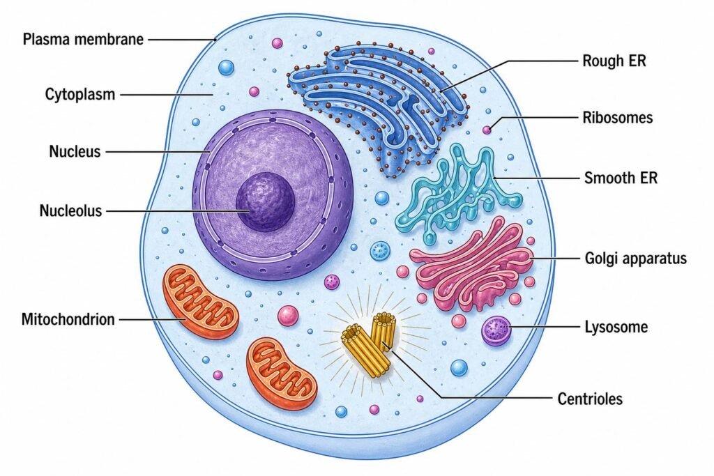
- Cellular organelles, including mitochondria, the endoplasmic reticulum, lysosomes, and the nucleus, perform specialized functions that contribute to normal cellular metabolism.
Definition of a Cell
- A cell is the smallest structural and functional unit of a living organism capable of performing essential life processes.
- It contains various biomolecules, enzymes, membranes, and specialized structures that work together to maintain cellular activities.
- The cell obtains nutrients, produces energy, synthesizes proteins, removes waste products, and responds to environmental signals.
- In humans, cells are organized into tissues, organs, and organ systems.
- Examples include hepatocytes in the liver, neurons in the nervous system, osteoblasts in bone, and erythrocytes in blood.
Types of Cells
Cells are broadly classified into two major types based on their structural organization.
A. Prokaryotic Cells
- Prokaryotic cells lack a membrane-bound nucleus.
- Their genetic material is located in a region called the nucleoid.
- They lack membrane-bound organelles such as mitochondria and the Golgi apparatus.
- They contain ribosomes that synthesize proteins.
- Most bacteria are examples of prokaryotic organisms.
Biochemical significance:
- Prokaryotic cells perform metabolic reactions using enzymes present in the cytoplasm or associated with the plasma membrane.
- Bacterial ribosomes differ structurally from human cytoplasmic ribosomes, which is important in understanding the selective action of certain antibiotics.
B. Eukaryotic Cells
- Eukaryotic cells possess a well-defined nucleus enclosed by a nuclear membrane.
- They contain membrane-bound organelles that perform specialized functions.
- Their DNA is organized into chromosomes within the nucleus.
- They contain mitochondria, endoplasmic reticulum, Golgi apparatus, lysosomes, and other cellular structures.
- Human cells are examples of eukaryotic cells.
Biochemical significance:
- Different metabolic pathways occur in specific cellular compartments.
- For example, glycolysis occurs in the cytosol, whereas the citric acid cycle occurs primarily in the mitochondrial matrix.
- This compartmentalization allows cells to regulate biochemical reactions efficiently.
Chemical Composition of the Cell
The human cell contains water, inorganic ions, and organic biomolecules that are essential for cellular structure and function.
1. Water
- Water is the most abundant chemical component of most living cells, generally accounting for approximately 60–70% of cell mass, although the proportion varies with cell type and physiological conditions.
- It acts as a solvent for numerous biochemical reactions.
- It provides a medium for the transport of nutrients, electrolytes, and metabolic waste products.
- It participates directly in biochemical reactions, including hydrolysis.
- It helps regulate cellular temperature and maintain cell volume.
- It facilitates interactions between enzymes and their substrates.
Biochemical importance: Water is essential for metabolic reactions, including digestion, ATP metabolism, and the maintenance of cellular homeostasis.
2. Proteins
- Proteins are organic macromolecules composed of amino acids linked by peptide bonds.
- They perform structural, catalytic, transport, regulatory, and defensive functions.
- Enzymes catalyze biochemical reactions and regulate metabolic pathways.
- Structural proteins help maintain cell shape and tissue organization.
- Membrane proteins function as receptors, channels, carriers, and enzymes.
- Certain proteins participate in cell signalling, immunity, and muscle contraction.
Examples:
- Enzymes: Hexokinase, lactate dehydrogenase.
- Structural proteins: Actin, tubulin.
- Transport proteins: Hemoglobin, membrane transporters.
- Regulatory proteins: Insulin, transcription factors.
- Defensive proteins: Immunoglobulins.
Biochemical importance: Proteins are essential for metabolism, cellular structure, signal transduction, and the regulation of physiological functions.
3. Lipids
- Lipids are a diverse group of organic compounds that are generally insoluble or poorly soluble in water.
- They form an important structural component of biological membranes.
- Phospholipids form the basic framework of the plasma membrane.
- Cholesterol helps regulate membrane fluidity and stability.
- Triglycerides serve as energy-storage molecules.
- Certain lipids act as precursors of steroid hormones and signalling molecules.
- Glycolipids contribute to cell recognition and membrane organization.
Examples:
- Phospholipids
- Cholesterol
- Triglycerides
- Glycolipids
- Sphingolipids
Biochemical importance: Lipids are essential for membrane structure, energy storage, intracellular signalling, and the synthesis of steroid hormones.
4. Carbohydrates
- Carbohydrates are organic compounds composed primarily of carbon, hydrogen, and oxygen.
- Glucose is an important energy source for many cells.
- Carbohydrates undergo glycolysis to generate ATP and metabolic intermediates.
- Glycogen stores glucose in the liver and skeletal muscles.
- Carbohydrate chains attached to proteins and lipids participate in cell recognition and adhesion.
- Carbohydrates also contribute to the structure of certain cellular molecules.
Examples:
- Monosaccharides: Glucose, fructose, ribose.
- Disaccharides: Sucrose, lactose, maltose.
- Polysaccharides: Glycogen.
Biochemical importance: Carbohydrates provide metabolic energy, supply intermediates for biosynthesis, and contribute to cellular recognition.
5. Nucleic Acids
Nucleic acids are biomolecules responsible for storing, transmitting, and expressing genetic information. They are composed of nucleotide units.
A. Deoxyribonucleic Acid (DNA)
- DNA stores hereditary information.
- It contains the instructions required for protein synthesis and cellular function.
- DNA replication ensures the transmission of genetic information to daughter cells.
- In human cells, most DNA is located in the nucleus, while mitochondria contain their own DNA.
B. Ribonucleic Acid (RNA)
- RNA participates in the expression of genetic information.
- Messenger RNA (mRNA) carries genetic information from DNA to ribosomes.
- Transfer RNA (tRNA) transports amino acids during protein synthesis.
- Ribosomal RNA (rRNA) forms an essential structural and catalytic component of ribosomes.
Biochemical importance: Nucleic acids regulate gene expression, protein synthesis, cellular growth, and inheritance.
6. Inorganic Ions and Minerals
- Inorganic ions are essential for maintaining cellular structure, electrical activity, enzyme function, and fluid balance.
- Sodium and potassium help establish membrane potential and regulate cellular volume.
- Calcium acts as an intracellular second messenger and participates in muscle contraction.
- Magnesium supports numerous enzyme reactions and interacts with ATP.
- Chloride contributes to electrical neutrality and fluid balance.
- Phosphate participates in ATP metabolism, phosphorylation reactions, and intracellular buffering.
- Iron is required for proteins involved in oxygen transport and electron transfer.
Examples and functions:
| Ion or mineral | Major biochemical function |
|---|---|
| Sodium (Na⁺) | Maintains extracellular fluid balance and contributes to membrane potential |
| Potassium (K⁺) | Maintains intracellular ionic balance and membrane potential |
| Calcium (Ca²⁺) | Cell signalling, muscle contraction, and secretion |
| Magnesium (Mg²⁺) | Enzyme activity and ATP-dependent reactions |
| Chloride (Cl⁻) | Ionic balance and electrical neutrality |
| Phosphate (PO₄³⁻) | ATP synthesis, phosphorylation, and buffering |
| Iron (Fe) | Oxygen transport and electron transfer |
Biochemical importance: Electrolytes and minerals are necessary for enzyme activity, energy metabolism, nerve transmission, and cellular homeostasis.
7. Vitamins and Coenzymes
- Vitamins are organic micronutrients required in small amounts for normal metabolic functions.
- Many B-complex vitamins act as precursors of coenzymes involved in energy metabolism.
- Coenzymes assist enzymes in transferring electrons, functional groups, or chemical groups during biochemical reactions.
- Vitamin-derived coenzymes participate in carbohydrate, lipid, amino acid, and nucleotide metabolism.
- Some vitamins, such as vitamins C and E, also contribute to antioxidant defence.
Examples:
- Vitamin B1: Precursor of thiamine pyrophosphate (TPP).
- Vitamin B2: Precursor of FAD and FMN.
- Vitamin B3: Precursor of NAD⁺ and NADP⁺.
- Vitamin B6: Precursor of pyridoxal phosphate (PLP).
- Vitamin B9: Precursor of tetrahydrofolate (THF).
- Vitamin B12: Required for reactions involving methylcobalamin and adenosylcobalamin.
- Vitamin C: Supports collagen synthesis and antioxidant functions.
Biochemical importance: Vitamins and their derived coenzymes support enzyme activity and maintain normal cellular metabolism.
8. Cellular Metabolites
- Metabolites are small molecules produced, consumed, or transformed during cellular biochemical reactions.
- They serve as substrates, intermediates, or products of metabolic pathways.
- Some metabolites participate in energy transfer, biosynthesis, and cellular signalling.
- Their concentrations change according to metabolic activity and physiological conditions.
Examples:
- Glucose
- Pyruvate
- Lactate
- Acetyl-CoA
- Amino acids
- Fatty acids
- ATP
- NADH
Biochemical importance: Metabolites connect different metabolic pathways and help regulate energy production, biosynthesis, and cellular responses.
Structure and Biochemical Functions of Cell Organelles
Cell organelles are specialized structures that perform distinct biochemical functions.
1. Plasma Membrane
Structure
- The plasma membrane is the outer boundary of the cell that separates the intracellular environment from the extracellular environment.
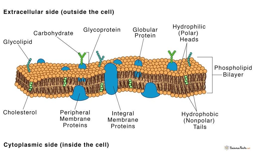
- It consists mainly of a phospholipid bilayer containing proteins, cholesterol, and carbohydrates.
- Phospholipids have hydrophilic heads facing the aqueous environments and hydrophobic tails directed towards the interior of the bilayer.
- Membrane proteins may be integral or peripheral and perform transport, receptor, enzymatic, and structural functions.
- Cholesterol helps regulate membrane fluidity and stability.
- Carbohydrates attached to proteins and lipids form part of the glycocalyx, which participates in cell recognition and adhesion.
Biochemical Functions
- Regulates the movement of ions, nutrients, and metabolic waste products across the cell membrane.
- Facilitates glucose and amino acid transport through specific carrier proteins.
- Maintains electrochemical gradients through membrane transport proteins and ion pumps.
- Contains receptors that recognize hormones, neurotransmitters, and other signalling molecules.
- Participates in signal transduction and the regulation of cellular metabolism.
- Supports endocytosis and exocytosis for the uptake and release of substances.
Clinical significance: Defects in membrane proteins can cause disorders involving ion transport, glucose uptake, and cellular communication.
2. Nucleus
Structure
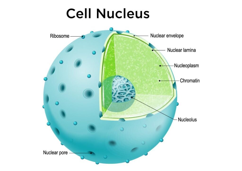
- The nucleus is a membrane-bound organelle that contains most of the genetic material of a eukaryotic cell.
- It is surrounded by a double membrane called the nuclear envelope.
- Nuclear pores regulate the movement of RNA, proteins, and other molecules between the nucleus and cytoplasm.
- The nucleoplasm contains chromatin, nucleotides, enzymes, and other molecules involved in genetic processes.
- Chromatin consists of DNA associated with proteins, particularly histones.
- The nucleolus is a specialized region involved in ribosomal RNA synthesis and ribosome assembly.
Biochemical Functions
- Stores and protects genetic information in DNA.
- Supports DNA replication before cell division.
- Provides the site for transcription of DNA into RNA.
- Regulates gene expression and the synthesis of cellular proteins.
- Participates in DNA repair and the maintenance of genomic integrity.
- Supports ribosomal RNA synthesis and the assembly of ribosomal subunits in the nucleolus.
Clinical significance: Mutations, DNA damage, and abnormalities in gene regulation can contribute to genetic disorders and cancer.
3. Mitochondria
Structure
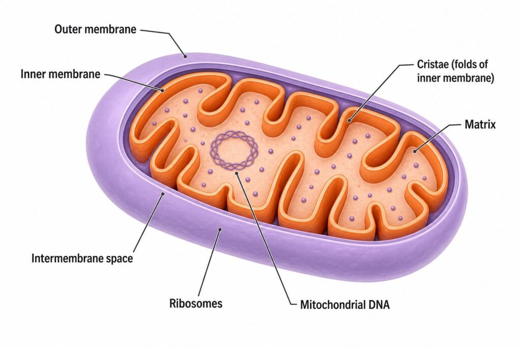
- Mitochondria are double-membrane-bound organelles responsible for major aspects of cellular energy metabolism.
- The outer membrane surrounds the organelle and contains proteins involved in molecular transport.
- The inner membrane is highly folded into structures called cristae, which increase the surface area available for energy production.
- The intermembrane space lies between the outer and inner membranes.
- The mitochondrial matrix contains enzymes, mitochondrial DNA, RNA, and ribosomes.
- The inner membrane contains the electron transport chain and ATP synthase.
Biochemical Functions
- Produces ATP through oxidative phosphorylation.
- Contains enzymes involved in the citric acid cycle.
- Participates in the beta-oxidation of fatty acids.
- Supports pyruvate oxidation and the conversion of pyruvate into acetyl-CoA.
- Participates in calcium regulation and cellular signalling.
- Plays an important role in the intrinsic pathway of apoptosis through the release of cytochrome c under appropriate conditions.
- Produces reactive oxygen species as by-products of cellular metabolism.
Clinical significance: Mitochondrial dysfunction can impair ATP production and contribute to neurological, muscular, and metabolic disorders.
4. Endoplasmic Reticulum
- The endoplasmic reticulum (ER) is a network of interconnected membranes involved in protein synthesis, lipid metabolism, and intracellular transport.
- It is classified into rough and smooth endoplasmic reticulum.
A. Rough Endoplasmic Reticulum (RER)
Structure
- Consists of flattened membrane-bound sacs called cisternae.
- Ribosomes are attached to its cytoplasmic surface, giving it a rough appearance.
- It is often continuous with the outer nuclear membrane.
Biochemical Functions
- Synthesizes proteins destined for secretion, insertion into cellular membranes, or delivery to certain organelles.
- Supports protein folding and quality control.
- Participates in the initial processing and glycosylation of many newly synthesized proteins.
- Transports newly synthesized proteins towards the Golgi apparatus.
- Helps prevent the accumulation of improperly folded proteins through cellular quality-control mechanisms.
Example: Plasma cells contain abundant rough endoplasmic reticulum because they synthesize and secrete large quantities of antibodies.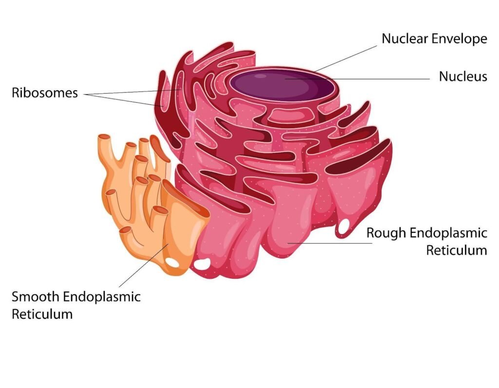
B. Smooth Endoplasmic Reticulum (SER)
Structure
- Consists of a network of membrane-bound tubules.
- Lacks ribosomes on its cytoplasmic surface.
- Its abundance varies according to the metabolic functions of the cell.
Biochemical Functions
- Participates in phospholipid and other lipid synthesis.
- Supports steroid hormone synthesis in steroid-producing cells.
- Contributes to drug and toxin metabolism, particularly in hepatocytes.
- Stores and releases calcium ions.
- Participates in glycogen metabolism indirectly through enzymes associated with its membranes in certain tissues.
Example: Smooth endoplasmic reticulum is prominent in steroid-producing cells and hepatocytes.
Clinical significance: Endoplasmic reticulum stress and abnormal protein folding are associated with several metabolic, genetic, and degenerative disorders.
5. Golgi Apparatus
Structure
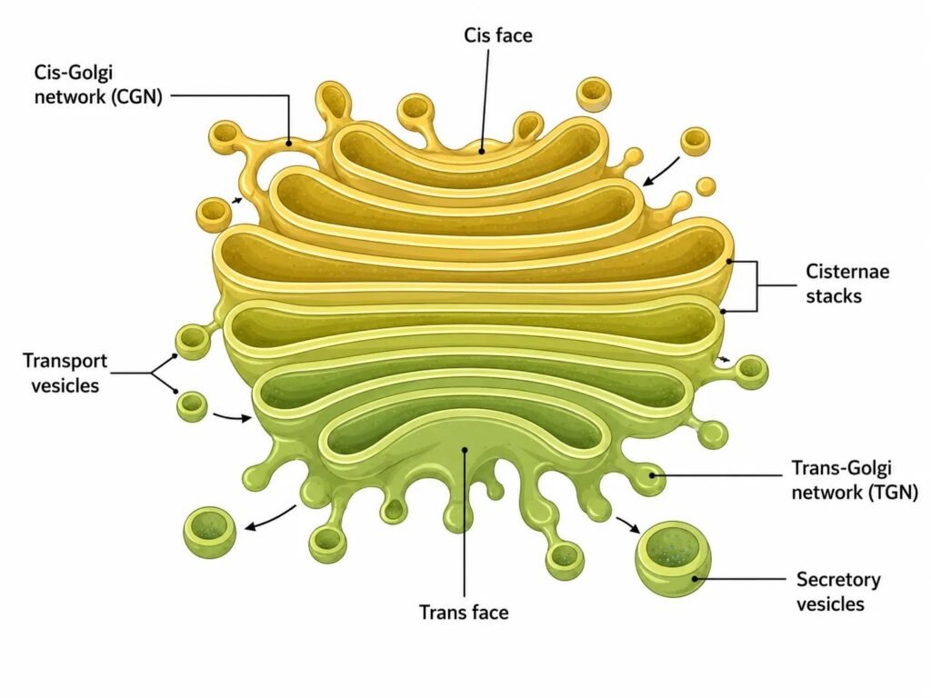
- The Golgi apparatus consists of stacks of flattened, membrane-bound sacs called cisternae.
- It has a cis face that receives materials from the endoplasmic reticulum and a trans face that directs processed materials towards their destinations.
- Transport vesicles move proteins and lipids between the ER, Golgi apparatus, and other cellular compartments.
Biochemical Functions
- Modifies proteins and lipids received from the endoplasmic reticulum.
- Performs glycosylation and other post-translational modifications.
- Sorts and packages proteins into transport or secretory vesicles.
- Directs selected enzymes towards lysosomes.
- Participates in the formation and renewal of cellular membranes.
- Supports the secretion of hormones, enzymes, and other cellular products.
Clinical significance: Abnormalities in Golgi-mediated protein processing and trafficking can interfere with normal cellular function.
6. Lysosomes
Structure
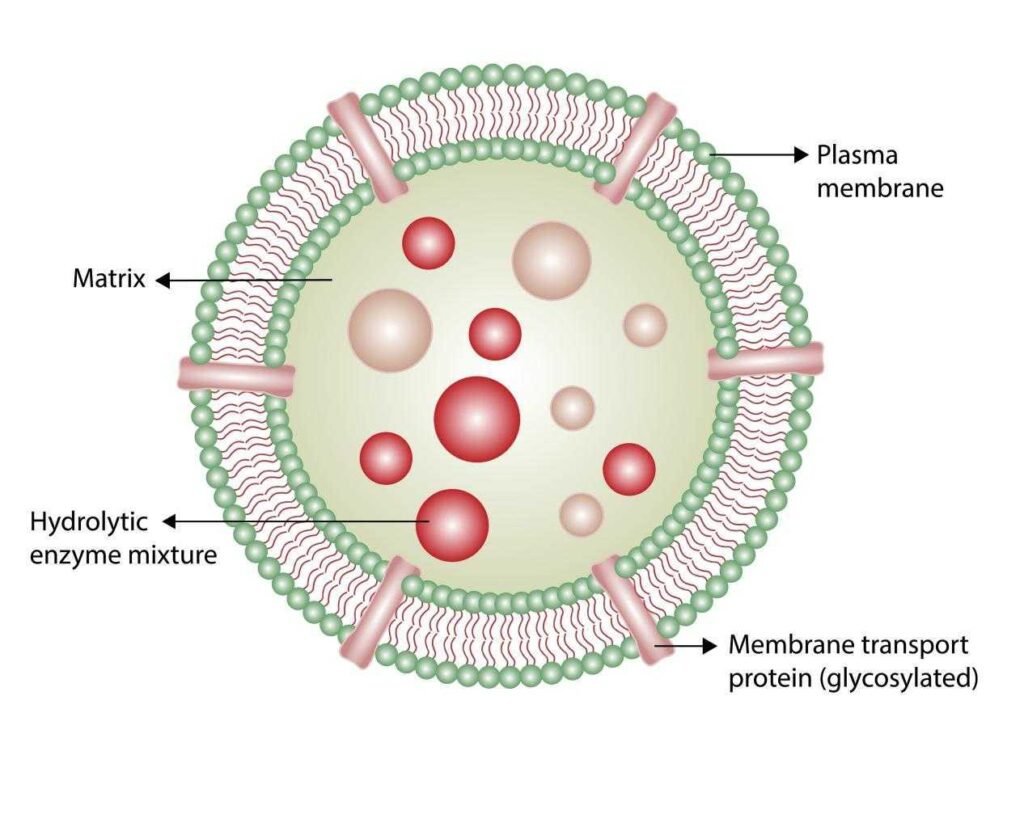
- Lysosomes are single-membrane-bound organelles containing digestive enzymes.
- Their enzymes include proteases, lipases, nucleases, glycosidases, and phosphatases.
- Lysosomal enzymes function optimally in an acidic environment maintained by proton pumps in the lysosomal membrane.
- The lysosomal membrane separates digestive enzymes from the cytoplasm.
Biochemical Functions
- Degrades proteins into smaller peptides and amino acids.
- Breaks down lipids, carbohydrates, and nucleic acids.
- Digests substances taken up through endocytosis and phagocytosis.
- Participates in autophagy, allowing cells to degrade and recycle damaged organelles.
- Recycles cellular components for reuse in biosynthetic pathways.
- Contributes to intracellular defence by digesting certain ingested microorganisms.
Clinical significance: Deficiency of specific lysosomal enzymes can cause lysosomal storage diseases.
Examples include:
- Gaucher disease: Deficiency of glucocerebrosidase.
- Tay-Sachs disease: Deficiency of hexosaminidase A.
- Pompe disease: Deficiency of acid alpha-glucosidase.
7. Peroxisomes
Structure
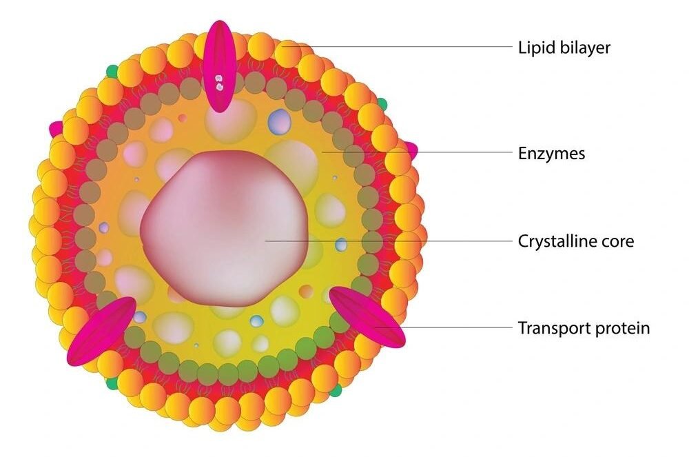
- Peroxisomes are small, single-membrane-bound organelles.
- They contain oxidative enzymes, including oxidases and catalase.
- Their enzyme composition varies according to the cell type and metabolic requirements.
Biochemical Functions
- Participate in the breakdown of very-long-chain fatty acids through beta-oxidation.
- Generate hydrogen peroxide during certain oxidative reactions.
- Contain catalase, which converts hydrogen peroxide into water and oxygen.
- Participate in the synthesis of plasmalogens, which are important membrane lipids, particularly in nervous tissue.
- Contribute to bile acid synthesis in the liver.
- Help maintain cellular lipid metabolism and redox balance.
Clinical significance: Peroxisomal dysfunction can cause severe metabolic disorders, including Zellweger spectrum disorders.
8. Ribosomes
Structure
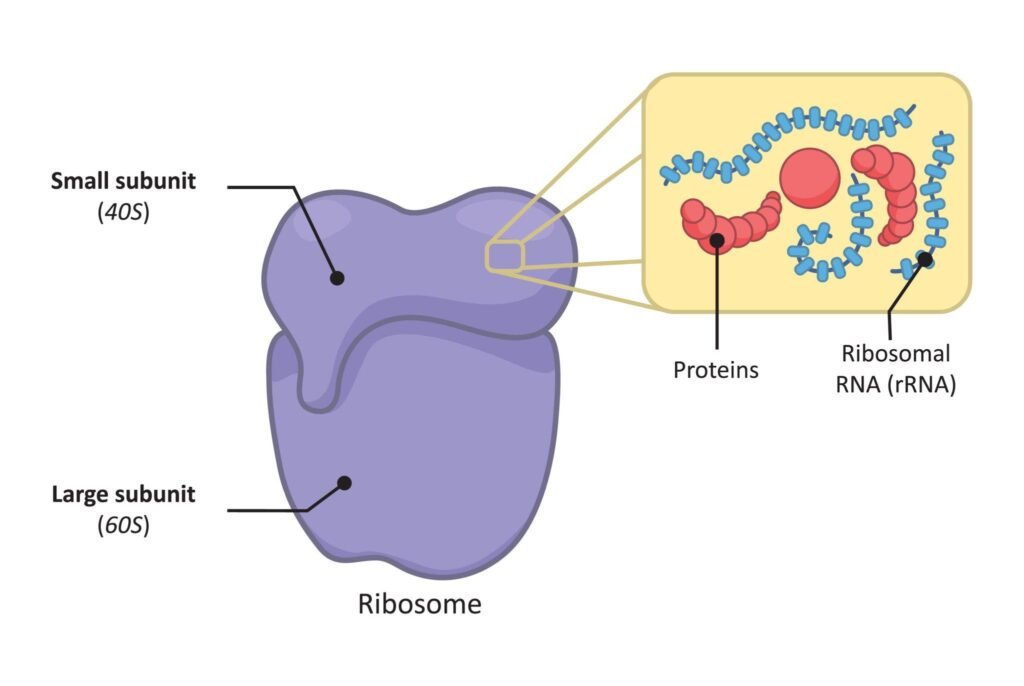
- Ribosomes are non-membrane-bound structures composed of ribosomal RNA and proteins.
- They consist of a small subunit and a large subunit.
- In human cells, cytoplasmic ribosomes are 80S ribosomes, consisting of 40S and 60S subunits.
- Mitochondria contain their own ribosomes, which differ from cytoplasmic ribosomes.
- Ribosomes may be free in the cytoplasm or attached to the rough endoplasmic reticulum.
Biochemical Functions
- Synthesizes proteins through the process of translation.
- Reads codons on messenger RNA to determine the sequence of amino acids.
- Facilitates the interaction of messenger RNA, transfer RNA, and amino acids.
- Catalyses peptide bond formation through the ribosomal RNA of the large subunit.
- Free ribosomes generally synthesize proteins used in the cytosol or targeted to certain organelles.
- ER-bound ribosomes synthesize many proteins destined for secretion, membranes, or the endomembrane system.
Clinical significance: Ribosomal abnormalities can impair protein synthesis and contribute to disorders known as ribosomopathies.
9. Centrosome and Centrioles
Structure
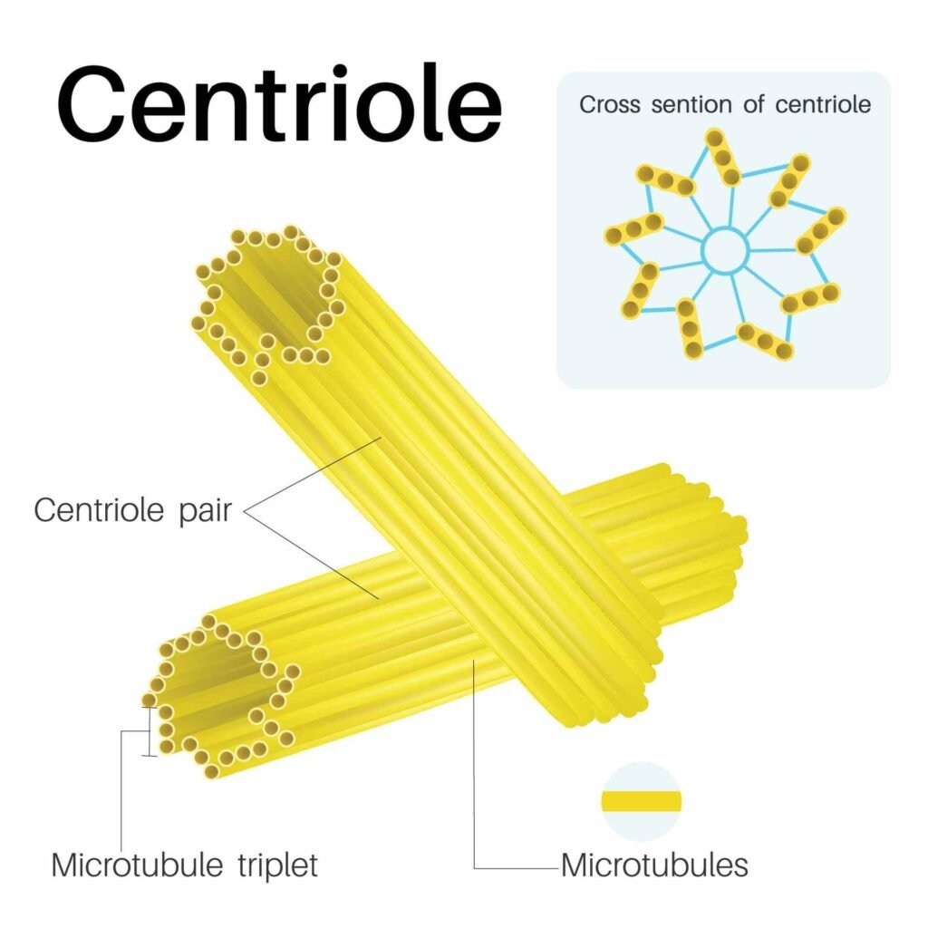
- The centrosome is a major microtubule-organizing centre in most animal cells.
- It typically contains two centrioles surrounded by pericentriolar material.
- Each centriole consists of nine microtubule triplets arranged in a cylindrical pattern.
Biochemical Functions
- Organizes microtubules within the cell.
- Helps establish the mitotic spindle during cell division.
- Contributes to the accurate separation of chromosomes during mitosis.
- Supports intracellular organization and the positioning of certain organelles.
- Centrioles can also contribute to the formation of basal bodies, which organize cilia and flagella.
Clinical significance: Abnormalities in centrosome function can contribute to chromosome segregation errors and abnormal cell proliferation.
10. Cytoskeleton
Structure
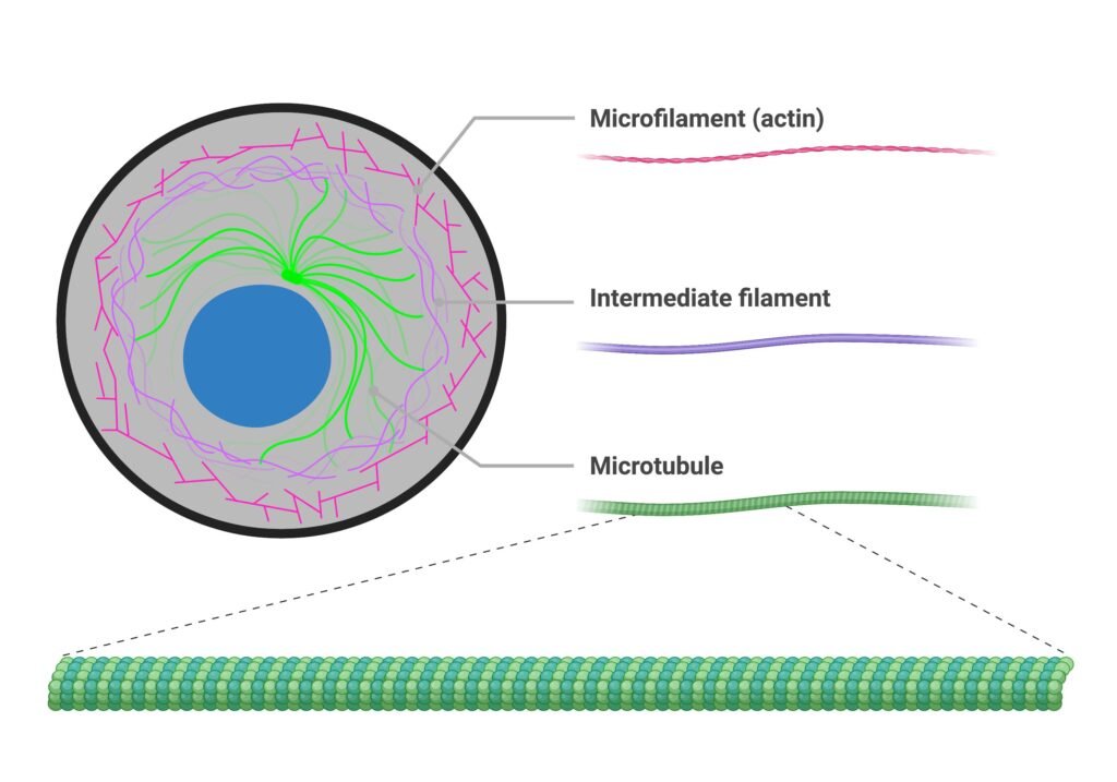
The cytoskeleton is a dynamic network of protein filaments distributed throughout the cytoplasm. It consists of three major components:
- Microfilaments: Primarily composed of actin.
- Intermediate filaments: Composed of different proteins, including keratins, vimentin, and lamins.
- Microtubules: Composed of alpha- and beta-tubulin.
Biochemical Functions
- Maintains cell shape and mechanical stability.
- Supports intracellular transport and organelle positioning.
- Participates in cell movement and changes in cell shape.
- Supports chromosome movement during cell division through microtubule-based structures.
- Actin and myosin interact during muscle contraction and other cellular movements.
- Microtubules provide tracks for motor proteins such as kinesin and dynein.
Clinical significance: Cytoskeletal defects can affect muscle function, intracellular transport, cell movement, and tissue integrity.
11. Cytoplasm and Cytosol
Structure
- The cytoplasm includes the cytosol, organelles, and other structures located between the plasma membrane and the nucleus.
- The cytosol is the aqueous intracellular fluid containing ions, proteins, metabolites, and soluble enzymes.
- Many biochemical reactions occur in the cytosol.
Biochemical Functions
- Provides the site for glycolysis.
- Supports several steps of amino acid and nucleotide metabolism.
- Contains enzymes involved in glycogen synthesis and breakdown.
- Facilitates intracellular movement of metabolites and other molecules.
- Supports protein synthesis by free ribosomes.
- Helps maintain the intracellular environment required for enzyme activity.
Clinical significance: Changes in cytosolic enzyme activity or metabolite concentrations can provide information about cellular metabolism and disease.
Cell Membrane
Structure of the Cell Membrane
- The cell membrane is approximately 7–10 nm thick, although its thickness varies depending on the cell type and membrane composition.
- It consists mainly of a phospholipid bilayer containing proteins, cholesterol, and carbohydrates.
- The hydrophilic portions of phospholipids face the aqueous environments inside and outside the cell.
- The hydrophobic fatty acid tails face inward, forming a barrier to many water-soluble substances.
- Membrane proteins are embedded within or associated with the lipid bilayer.
- Cholesterol helps regulate membrane fluidity, stability, and permeability.
- Carbohydrates attached to proteins and lipids contribute to the glycocalyx, which is involved in cell recognition and adhesion.
Fluid Mosaic Model
The fluid mosaic model, proposed by Singer and Nicolson in 1972, describes the plasma membrane as a dynamic structure in which proteins are distributed within a fluid lipid bilayer.
Major Features
- Fluid nature: Many membrane lipids and proteins can move laterally within the membrane.
- Mosaic arrangement: Different proteins are distributed throughout the lipid bilayer.
- Selective permeability: The membrane permits certain substances to cross more easily than others.
- Membrane asymmetry: The inner and outer leaflets differ in their lipid and carbohydrate composition.
- Dynamic organization: Membrane components can reorganize in response to cellular signals and physiological requirements.
Biochemical importance: Membrane fluidity and the distribution of membrane proteins influence transport, receptor activity, cell signalling, and interactions with other cells.
Transport Across the Cell Membrane
The plasma membrane regulates the movement of substances between the intracellular and extracellular environments.
Types of Transport Across the Cell Membrane
Transport across the cell membrane is broadly classified into three major types:
- Passive transport: Movement of substances without direct energy expenditure by the cell.
- Active transport: Movement of substances using energy, often against their concentration or electrochemical gradient.
- Vesicular transport: Movement of large particles or molecules through membrane-bound vesicles.
1. Passive Transport
- Passive transport is the movement of substances across the cell membrane without direct expenditure of cellular energy in the form of ATP.
- Substances generally move down their concentration or electrochemical gradients.
A. Simple Diffusion
- Simple diffusion is the movement of molecules from a region of higher concentration to a region of lower concentration.
- It occurs directly through the phospholipid bilayer without the assistance of carrier or channel proteins.
- It is mainly used by small, non-polar, lipid-soluble molecules.
- Examples include oxygen (O₂), carbon dioxide (CO₂), and some steroid hormones.
- The rate of diffusion depends on the concentration gradient, membrane permeability, temperature, and molecular properties.
Biochemical importance: Simple diffusion allows oxygen to enter cells for cellular respiration and carbon dioxide to leave cells as a metabolic waste product.
B. Facilitated Diffusion
- Facilitated diffusion is the movement of substances across the cell membrane with the help of specific membrane proteins.
- It occurs down the concentration or electrochemical gradient and does not require direct ATP expenditure.
- It involves two major types of transport proteins: channel proteins and carrier proteins.
- Channel proteins form selective pathways for ions or water.
- Carrier proteins bind specific substances and undergo conformational changes to transport them across the membrane.
- This process is selective and may become saturated when all available transport proteins are occupied.
Examples:
- Glucose transport into many cells through GLUT transporters.
- Movement of potassium (K⁺), sodium (Na⁺), and chloride (Cl⁻) ions through appropriate ion channels.
- Water movement through aquaporins.
Biochemical importance: Facilitated diffusion supports glucose uptake, ion movement, and the maintenance of normal cellular function.
C. Osmosis
- Osmosis is the net movement of water across a selectively permeable membrane in response to differences in water potential.
- Water generally moves from a region of higher water potential to a region of lower water potential.
- In biological systems, the movement of water is influenced by the concentration of osmotically active solutes.
- Water can cross the membrane directly, but aquaporin channels facilitate rapid water movement in many cell types.
Types of solutions based on tonicity:
- Isotonic solution: Has the same effective osmotic concentration as the cell; the cell generally maintains its normal volume.
- Hypotonic solution: Has a lower effective osmotic concentration than the cell; water enters the cell, which may swell.
- Hypertonic solution: Has a higher effective osmotic concentration than the cell; water leaves the cell, which may shrink.
Clinical significance: Osmosis is important in maintaining cell volume and understanding the effects of intravenous fluids on red blood cells and other tissues.
D. Filtration
- Filtration is the movement of water and small dissolved substances across a membrane due to a pressure difference.
- It is driven mainly by hydrostatic pressure rather than directly by ATP.
- The movement of substances depends on membrane permeability, pore size, and the pressure gradient.
- It is particularly important in the movement of fluid across capillary walls and in the kidneys.
Example: Glomerular filtration in the kidneys allows water and small solutes to pass from blood into the glomerular filtrate.
2. Active Transport
- Active transport is the movement of substances across the cell membrane using energy.
- It can move substances against their concentration or electrochemical gradients.
Active transport is classified into primary and secondary active transport.
A. Primary Active Transport
- Primary active transport uses energy directly from ATP hydrolysis to move substances across the membrane.
- It is carried out by specialised membrane proteins called ATP-driven pumps.
- These pumps maintain essential ionic gradients and support cellular homeostasis.
Important examples:
1. Sodium-Potassium ATPase (Na⁺/K⁺-ATPase)
- It is present in the plasma membrane of most animal cells.
- For each ATP molecule hydrolysed, it transports three Na⁺ ions out of the cell and two K⁺ ions into the cell.
- It helps maintain intracellular potassium and low intracellular sodium concentrations.
- It contributes to the resting membrane potential and supports secondary active transport.
2. Calcium ATPase (Ca²⁺-ATPase)
- It transports calcium ions away from the cytosol.
- It helps maintain low cytosolic calcium concentrations.
- It is important in muscle contraction, cell signalling, and intracellular calcium regulation.
3. Proton Pumps (H⁺-ATPases)
- They transport hydrogen ions across membranes.
- They help establish acidic environments in certain cellular compartments.
- They are important for lysosomal acidification and other cellular processes.
Biochemical importance: Primary active transport maintains ionic gradients required for nerve impulse transmission, muscle function, nutrient transport, and cellular metabolism.
B. Secondary Active Transport
- Secondary active transport uses the energy stored in an ion gradient rather than directly hydrolysing ATP at the transporter itself.
- The ion gradient is usually established by primary active transport, such as the Na⁺/K⁺-ATPase.
- As an ion moves down its electrochemical gradient, another substance is transported against its own gradient.
- It is classified into symport and antiport.
1. Symport (Co-transport)
- Two or more substances move across the membrane in the same direction.
- The movement of one substance down its gradient drives the transport of another substance.
Example: Sodium-Glucose Linked Transporter (SGLT)
- Sodium ions and glucose move into the cell in the same direction.
- The inward movement of sodium provides the energy needed to transport glucose against its concentration gradient.
- SGLT1 is important in intestinal glucose absorption and renal glucose reabsorption, while SGLT2 plays a major role in glucose reabsorption in the proximal renal tubule.
2. Antiport (Counter-transport)
- Two substances move across the membrane in opposite directions.
- The movement of one substance down its gradient helps drive the movement of another substance in the opposite direction.
Examples:
- Sodium-calcium exchanger (NCX), which commonly uses the inward sodium gradient to move calcium out of cells.
- Sodium-hydrogen exchanger (NHE), which commonly uses the sodium gradient to transport hydrogen ions out of cells.
Biochemical importance: Secondary active transport is essential for nutrient absorption, renal tubular reabsorption, pH regulation, and calcium homeostasis.
3. Vesicular Transport
- Vesicular transport involves the movement of large molecules, particles, or bulk material using membrane-bound vesicles.
- It requires cellular energy and coordinated membrane remodelling.
A. Endocytosis
- Endocytosis is the process by which cells take substances into the cell through the formation of membrane-bound vesicles.
- The plasma membrane surrounds the material and forms a vesicle that enters the cell.
- It is important for nutrient uptake, immune defence, and receptor regulation.
Types of endocytosis:
1. Phagocytosis
- Phagocytosis is the uptake of large particles, microorganisms, or cellular debris.
- The cell surrounds the material and encloses it within a phagosome.
- The phagosome may fuse with a lysosome, allowing enzymatic digestion of the material.
- It is mainly performed by specialised cells such as macrophages and neutrophils.
Example: Engulfment of bacteria by macrophages.
2. Pinocytosis
- Pinocytosis is the uptake of extracellular fluid and dissolved substances through small vesicles.
- It allows cells to internalise fluid-phase material from their surroundings.
Example: Uptake of extracellular fluid and dissolved molecules by many cell types.
3. Receptor-Mediated Endocytosis
- This is a selective process in which specific substances bind to receptors on the cell surface.
- The receptor-ligand complexes are internalised in vesicles.
- It allows cells to take up particular molecules efficiently.
Example: Cellular uptake of LDL cholesterol through LDL receptors.
B. Exocytosis
- Exocytosis is the process by which intracellular vesicles fuse with the plasma membrane and release their contents outside the cell.
- It is important for secretion, waste removal, and the delivery of membrane proteins and lipids to the plasma membrane.
- Many forms of regulated exocytosis depend on calcium signalling.
Examples:
- Release of insulin from pancreatic beta cells.
- Secretion of neurotransmitters from nerve cells.
- Release of digestive enzymes from pancreatic acinar cells.
Biochemical importance: Exocytosis plays a major role in hormone secretion, neurotransmission, and the release of enzymes and other cellular products.
Comparison of Transport Mechanisms
| Feature | Simple Diffusion | Facilitated Diffusion | Active Transport | Vesicular Transport |
|---|---|---|---|---|
| Energy requirement | No direct ATP required | No direct ATP required | Energy required | Energy required |
| Transport proteins | Not required | Channels or carriers | Pumps or coupled transporters | Vesicle-forming and trafficking machinery |
| Direction | Down the concentration gradient | Down the concentration or electrochemical gradient | Can occur against a gradient | Depends on the process |
| Typical substances | O₂, CO₂ | Glucose, ions, water through aquaporins | Na⁺, K⁺, Ca²⁺, glucose in secondary transport | Proteins, large particles, bulk material |
| Examples | Oxygen entry into cells | Glucose transport through GLUT | Na⁺/K⁺-ATPase, SGLT | Endocytosis and exocytosis |
Factors Affecting Transport Across the Cell Membrane
The rate and direction of membrane transport depend on several factors:
- Concentration gradient: A larger concentration difference generally increases the rate of passive diffusion.
- Electrochemical gradient: Both concentration and electrical differences influence the movement of ions.
- Molecular size: Smaller molecules generally cross membranes more easily than larger molecules.
- Lipid solubility: Lipid-soluble molecules usually pass more readily through the phospholipid bilayer.
- Membrane permeability: The composition and structure of the membrane influence the movement of substances.
- Temperature: Temperature can affect molecular movement and membrane fluidity.
- Availability of transport proteins: The number and activity of channels, carriers, and pumps influence transport rates.
- ATP availability: Energy-dependent transport may be impaired when cellular ATP production decreases.
- Osmotic and hydrostatic pressure: These influence water movement and filtration across biological membranes.