Introduction
- Recombinant DNA technology is an important part of modern biotechnology and genetic engineering.
- It allows scientists to combine DNA fragments from different sources to produce a new DNA molecule.
- The process involves the isolation and manipulation of DNA under laboratory conditions.
- Recombinant DNA can contain genetic material obtained from different organisms or different sources.
- The newly formed DNA molecule can be introduced into a suitable host cell, where it can be replicated.
- When the inserted gene is expressed, the host cell can produce the protein corresponding to that gene.
- Important tools used in recombinant DNA technology include restriction enzymes, vectors, and DNA ligase.
- Recombinant DNA technology is closely associated with gene cloning, which allows a large number of identical copies of a selected DNA sequence to be produced.
- The technology has important applications in medicine, disease diagnosis, genetic research, agriculture, forensic science, and industry.
- It has also made possible the production of useful biological products such as insulin, therapeutic proteins, blood-clotting factors, and recombinant vaccines.
Recombinant DNA
- Recombinant DNA (rDNA) is DNA formed by combining two DNA fragments obtained from different sources.
- Genetic recombination is the natural exchange or combination of genetic material from different sources. It produces new combinations of genes.
- In natural genetic recombination, the newly formed genetic material can be replicated, transcribed, and translated.
- In eukaryotes, genetic recombination occurs during sexual reproduction. The chromosomes from the egg and sperm contribute genetic material to the offspring.
- During this process, corresponding parts of chromosomes can be exchanged and joined together, producing new combinations of genes.
Artificial Recombinant DNA
- Genes or groups of genes can also be combined artificially in vitro, meaning under laboratory conditions.
- Artificial recombinant DNA technology allows scientists to combine DNA fragments that may not normally occur together in nature.
- The process involves the isolation and manipulation of DNA to produce a hybrid or chimeric DNA molecule.
Chimeric DNA
- Chimeric DNA is a recombinant DNA molecule containing genetic material from two different species.
- For example, a chimeric DNA molecule may contain both human and bacterial DNA sequences.
- The term chimeric comes from the mythical creature Chimera, which was described as having the head of a lion, the body of a goat, and the tail of a snake.
Construction of Recombinant DNA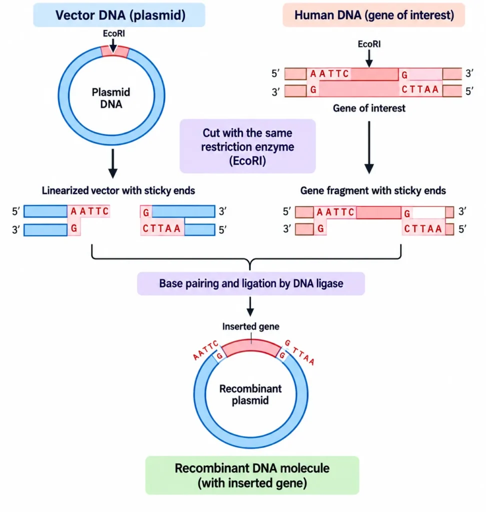
The basic steps involved are:
- Cutting of DNA
DNA is cut into fragments using restriction endonuclease enzymes. - Isolation of the desired DNA
The specific DNA fragment required for the experiment is separated from the other DNA fragments. - Insertion into a vector
The desired DNA fragment is inserted into a suitable vector or carrier DNA. - Joining of DNA fragments
The DNA fragment and vector are joined together using the enzyme DNA ligase.
Restriction Endonucleases
- Restriction endonucleases are also called restriction enzymes.
- They are found in many prokaryotic organisms.
- These enzymes recognize particular DNA sequences and cut DNA at specific sites.
- This specific cutting makes them useful for producing DNA fragments during recombinant DNA construction.
- Restriction enzymes are named according to the bacteria from which they are obtained. Examples include EcoRI from Escherichia coli and BamHI from Bacillus species.
- Most restriction enzymes recognize specific sequences containing about 4–6 base pairs.
- Many of these recognition sequences are palindromic, meaning the sequences have the same reading pattern in the 5′ to 3′ direction on both DNA strands.
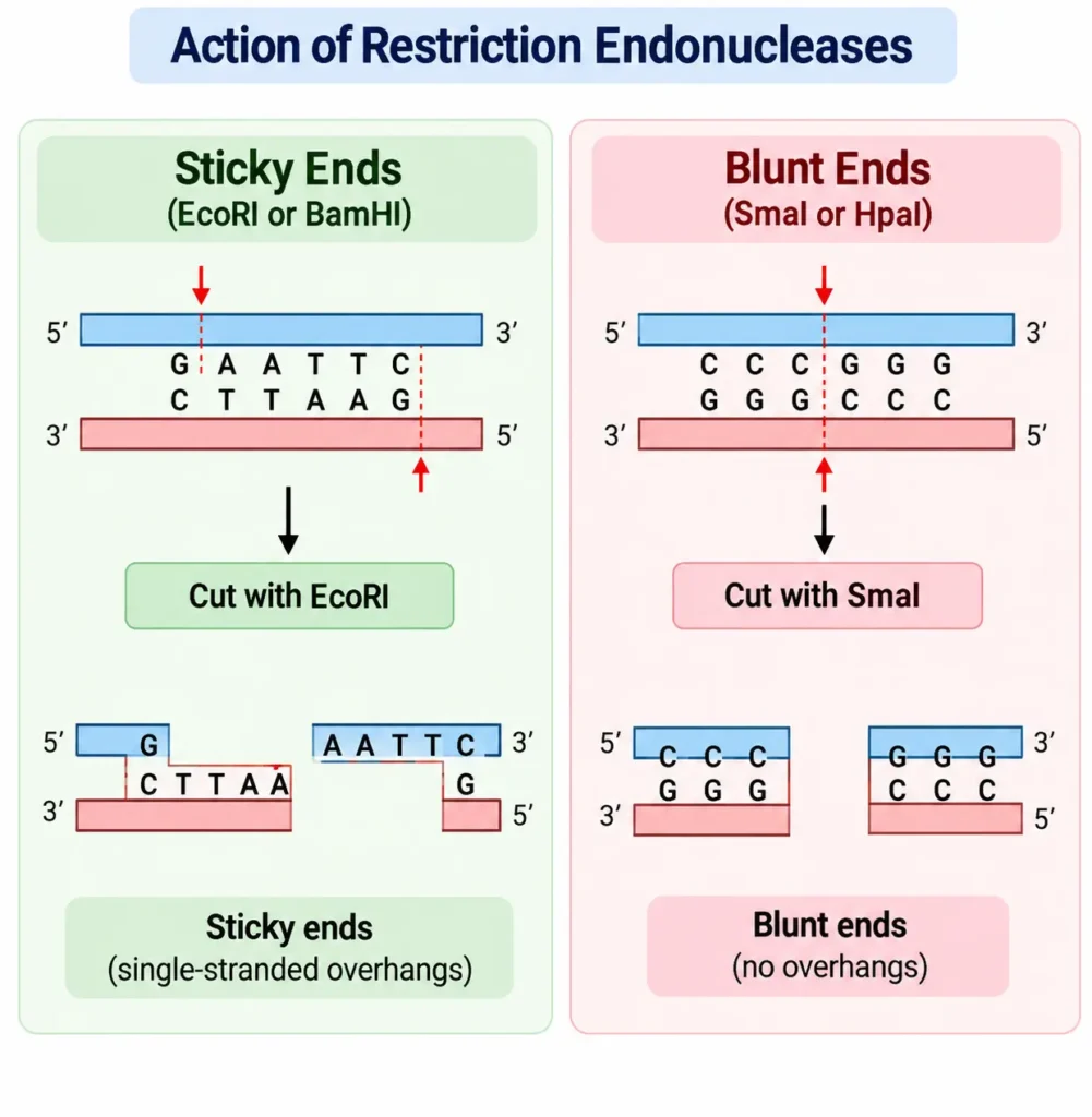
Sticky Ends and Blunt Ends
Restriction enzymes can produce two types of DNA ends:
- Blunt ends: Both DNA strands are cut at the same position, leaving no unpaired bases.
- Sticky ends: The two DNA strands are cut at different positions, leaving a short single-stranded region at the ends.
- Sticky ends are also called cohesive ends.
- Complementary sticky ends from DNA fragments cut by the same restriction enzyme can base-pair with each other.
- This property helps in joining DNA fragments from different sources.
Isolation of Specific DNA
- When DNA is cut by restriction enzymes, many different fragments are produced.
- The required DNA fragment must be separated from these fragments.
- Electrophoresis or HPLC can be used to separate the desired DNA fragment.
Insertion of DNA into a Vector
- A vector is a carrier DNA molecule used to carry the DNA fragment of interest.
- Foreign DNA usually cannot replicate independently inside a host cell.
- Therefore, the desired DNA fragment is joined to a vector that can replicate inside the host cell.
- Common cloning vectors include:
- Bacterial plasmids
- Bacteriophages
- Cosmids
Joining DNA by DNA Ligase
- After the DNA fragment and vector are brought together, complementary sticky ends can base-pair.
- The enzyme DNA ligase then joins their ends by forming covalent bonds.
- The final product is a recombinant, hybrid, or chimeric DNA molecule.
- Therefore, restriction enzymes and DNA ligase have important and complementary roles:
- Restriction enzymes → cut DNA
- DNA ligase → joins DNA fragments
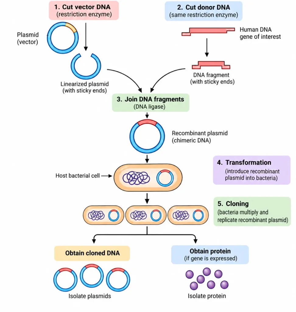
Cloning
- Cloning is a technique used to amplify DNA in vivo, meaning inside a living host cell.
- The main purpose of cloning is to produce a large number of identical copies of a particular DNA molecule.
- In recombinant DNA technology, the recombinant DNA is introduced into a suitable host cell, commonly a bacterial cell.
- The introduction of recombinant DNA into a bacterial host cell is called transformation.
- Host cells that contain the recombinant DNA are called transformed cells.
- The transformed cells are grown under suitable conditions that allow them to multiply rapidly.
- As the host cells divide, they replicate their own DNA as well as the vector containing the inserted DNA fragment.
- Repeated cell division therefore produces many copies of the recombinant DNA.
- The recombinant DNA can subsequently be isolated from the host cells in relatively large quantities.
- If the inserted gene is expressed under suitable conditions, the host cells can also produce the protein encoded by the inserted gene, which can then be isolated.
Applications of Recombinant DNA Technology
Recombinant DNA technology has many important applications in medicine, diagnosis, genetic research, agriculture, forensic science, and industry.
1. Production of Therapeutic Proteins
- Recombinant DNA technology is used to produce important human proteins in large quantities.
- Examples include:
- Insulin – used in the treatment of diabetes.
- Human growth hormone – used in children with growth hormone deficiency.
- Erythropoietin – used in the treatment of anemia.
- Factor VIII – used to treat patients with hemophilia.
- Tissue plasminogen activator (tPA) – used as an anticoagulant in the treatment of heart attack.
- Interferons – used in the treatment of certain cancers and other diseases.
- Interleukins – used in areas such as wound healing, cancer, and immune disorders.
- Monoclonal antibodies – used in diagnostic tests.
- Superoxide dismutase – has applications in medical treatment.
2. Vaccine Production
- Recombinant DNA technology can be used to produce vaccines.
- Recombinant hepatitis B vaccine is an important example of a vaccine produced using recombinant DNA technology.
3. Understanding Diseases
- Recombinant DNA technology helps in understanding the molecular basis of genetic diseases.
- It has been used in studying diseases such as:
- Familial hypercholesterolemia
- Sickle cell disease
- Thalassemia
- Cystic fibrosis
- Muscular dystrophy
4. Diagnosis of Diseases
- Recombinant DNA techniques can be used to identify and diagnose diseases.
- They are particularly useful in the study and detection of genetic disorders.
5. Gene Therapy
- Gene therapy involves introducing a normal gene into an individual who has a defective gene.
- Recombinant DNA technology provides an important basis for developing gene therapy approaches for genetic disorders such as sickle cell disease, thalassemia, and adenosine deaminase deficiency.
6. Transgenic Animals
- Genes can be introduced into fertilized eggs to produce transgenic animals.
- A transgenic animal contains genetic material that has been artificially introduced from another organism.
7. Genetic Counseling
- Recombinant DNA-based screening can help identify certain genetic defects in prospective parents.
- Fetal testing can also be performed to detect genetic abnormalities at an early stage.
8. Forensic Medicine
- DNA-based techniques such as DNA fingerprinting are used in forensic investigations.
- They can help establish an individual’s unique DNA pattern and may be useful in criminal cases.
9. Agricultural Applications
- Recombinant DNA technology is used to develop plants with useful characteristics.
- These include resistance to:
- Diseases
- Insects
- Herbicides
- Drought
- Extreme temperatures
- It can also be used in developing plants with improved nitrogen-fixing ability.
10. Industrial Applications
- Recombinant DNA technology is used in industrial biotechnology for the production of enzymes.
- These enzymes have applications in industries such as detergent, sugar, and cheese production.
Blot Transfer Techniques
- Blot transfer is a molecular biology technique used to identify and study specific DNA, RNA, or protein molecules from a mixture containing many different molecules.
- The three major types of blotting techniques are:
- Southern blot – used for DNA
- Northern blot – used for RNA
- Western blot – used for proteins
Southern Blot
Principle
- The principle of Southern blotting is based on the separation of DNA fragments according to size followed by specific hybridization between a DNA fragment and a complementary labeled probe.
- DNA is first cut into fragments and separated by gel electrophoresis.
- The DNA is then transferred to a membrane and exposed to a labeled cDNA probe.
- The probe hybridizes with the complementary DNA sequence, allowing the specific DNA fragment to be detected.
Procedure
1. DNA Extraction
- DNA is first extracted from the cells or tissue being studied.
- The extracted DNA contains many different DNA sequences.
2. Restriction Enzyme Digestion
- The DNA is treated with a suitable restriction enzyme.
- The enzyme cuts the DNA at its specific recognition sites.
- This produces many DNA fragments of different lengths.
3. Gel Electrophoresis
- The DNA fragments are separated by agarose or polyacrylamide gel electrophoresis.
- DNA fragments move through the gel according to their size.
- Smaller fragments move faster, while larger fragments move more slowly.
4. DNA Denaturation
- After electrophoresis, the DNA is treated with mild alkali.
- This denatures the double-stranded DNA into single strands.
- Single-stranded DNA is required for subsequent probe hybridization.
5. Transfer to Membrane
- The denatured DNA is transferred from the gel onto a nitrocellulose membrane.
- The transfer produces a replica of the DNA fragment pattern present in the gel.
- This transfer step is called blotting.
6. Probe Hybridization
- The membrane is exposed to a labeled cDNA probe.
- The probe recognizes and binds to the complementary DNA sequence on the membrane.
- This process is called hybridization.
7. Detection
- The membrane is examined to detect the labeled probe.
- In the procedure described in the book, the membrane is exposed to X-ray film.
- A specific band appears corresponding to the DNA fragment recognized by the probe.
Result
- A specific band or bands on the membrane indicate the presence of the DNA sequence complementary to the probe.
- The position of the band indicates the approximate size of the corresponding DNA fragment.
- The pattern obtained can therefore be used to identify specific DNA sequences.
Applications
Southern blotting can be used for:
- Identification of specific DNA sequences
- Determination of the number of copies of a gene in a tissue
- Detection or study of gene mutations
- Analysis of particular DNA fragments after restriction enzyme digestion
Northern Blot
Principle
- The principle of Northern blotting is based on the separation of RNA molecules according to their size followed by hybridization with a complementary labeled probe.
- The RNA is first separated by electrophoresis and then transferred to a membrane.
- A complementary probe binds to the specific RNA sequence of interest.
- This allows the particular RNA molecule to be identified.
Procedure
1. RNA Extraction
- RNA is first isolated from the cells or tissue being studied.
- The sample contains different types and sizes of RNA molecules.
2. Gel Electrophoresis
- The extracted RNA is subjected to gel electrophoresis.
- RNA molecules are separated according to their size.
- Smaller RNA molecules generally migrate faster through the gel than larger molecules.
3. Transfer to Membrane
- After electrophoresis, the separated RNA molecules are transferred from the gel onto a suitable membrane.
- The transfer preserves the pattern of RNA molecules obtained during electrophoresis.
4. Probe Hybridization
- The membrane is treated with a labeled complementary nucleic acid probe.
- The probe binds specifically to the complementary RNA sequence.
- This process is called hybridization.
5. Detection
- The hybrid formed between the RNA and complementary probe is detected using an appropriate detection method.
- In the procedure described in the source, the hybrids can be identified by radioautography.
Important Difference from Southern Blot
- In Southern blotting, DNA is analyzed.
- In Northern blotting, RNA is analyzed.
- An important difference in the procedure is that alkali is not used in Northern blotting because alkali can hydrolyze RNA.
Result
- A specific band or signal indicates the presence of the RNA molecule complementary to the probe.
- The position of the band provides information about the size of the RNA molecule.
- The intensity of the detected signal can provide information about the amount of the specific RNA present.
Applications
Northern blotting is useful for:
- Identification of specific RNA molecules
- Determination of the size of RNA
- Measurement of specific RNA molecules
- Study of gene expression by examining the RNA produced from a gene
Western Blot
Principle
- The principle of Western blotting is based on the separation of proteins according to their size followed by specific detection using antibodies.
- Proteins are first separated by electrophoresis and then transferred to a membrane.
- The membrane is treated with antibodies that specifically recognize the protein of interest.
- The labeled antibody allows the specific protein to be detected.
Procedure
1. Protein Extraction
- Proteins are extracted from the cells or tissue being studied.
- The sample contains many different proteins.
2. Gel Electrophoresis
- The proteins are separated by electrophoresis.
- Different proteins migrate through the gel according to their size.
3. Transfer to Membrane
- After electrophoresis, the separated proteins are transferred from the gel onto a nitrocellulose membrane.
- The protein pattern on the membrane corresponds to the pattern obtained in the gel.
4. Antibody Detection
- The membrane is treated with a specific labeled antibody.
- The antibody recognizes and binds to the particular protein of interest.
- This antibody-protein interaction provides the specificity of the technique.
5. Detection
- The labeled antibody is detected using an appropriate detection method.
- A visible band or signal indicates the presence of the specific protein.
Result
- A specific band indicates that the protein recognized by the antibody is present in the sample.
- The position of the band can provide information about the protein’s size.
- The amount of protein can be estimated from the strength of the detected signal.
Applications
Western blotting is used for:
- Identification of specific proteins
- Determination of the size of specific proteins
- Measurement of specific protein molecules
- Detection of proteins associated with particular diseases
- Research and diagnostic applications
Restriction Fragment Length Polymorphism
- Restriction Fragment Length Polymorphism (RFLP) is a molecular biology technique based on differences in DNA sequences that produce different lengths of restriction fragments.
- These differences can be used to identify genetic variation and certain genetic defects.
Principle
- DNA sequences can vary between individuals. Such variations are called polymorphisms.
- A variation in DNA sequence may create or eliminate a restriction enzyme recognition site.
- When DNA is cut with a restriction enzyme, these differences can produce DNA fragments of different lengths.
- The inherited difference in the pattern of restriction fragments is called Restriction Fragment Length Polymorphism (RFLP).
Procedure
The basic procedure can be understood as follows:
- DNA isolation – DNA is obtained from the sample.
- Restriction enzyme digestion – The DNA is cut using a specific restriction enzyme.
- Fragment separation – The resulting DNA fragments are separated according to their size, usually by gel electrophoresis.
- Pattern analysis – The resulting fragment pattern is compared between samples.
- Identification of variation – Differences in fragment patterns indicate differences in the DNA restriction sites.
Note: The provided book explains the principle and applications of RFLP but does not describe a detailed laboratory procedure. The above sequence is a simple conceptual representation of how RFLP analysis is performed.
Result
- RFLP produces a characteristic pattern of DNA fragments.
- If a DNA sequence variation changes a restriction site, the resulting fragments will have different lengths.
- Therefore, different individuals or samples may show different fragment patterns.
Applications
RFLP can be used for:
- Detection of genetic defects
- Study of inherited DNA variations
- Identification of genetic differences between individuals
- Analysis of genetic disorders such as:
- Sickle cell anemia
- Thalassemia
Polymerase Chain Reaction
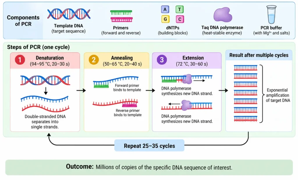
- Polymerase Chain Reaction (PCR) is an in-vitro technique used to amplify a specific DNA sequence.
- It can produce a large number of copies of a selected DNA region from a very small amount of starting DNA.
- PCR is useful in clinical testing, forensic investigations, microbiology, and molecular biology research.
Principle
- The principle of PCR is based on the repeated separation of DNA strands, binding of primers, and synthesis of new DNA strands.
- Each cycle produces additional copies of the target DNA.
- Repeating the cycle many times results in a large number of copies of the desired DNA sequence.
Components Required for PCR
The main components are:
- Template DNA – contains the DNA sequence to be amplified.
- Primers – short synthetic DNA sequences that bind to the regions flanking the target DNA.
- Deoxyribonucleotides (dNTPs) – building blocks used to synthesize new DNA.
- DNA polymerase – enzyme responsible for synthesizing new DNA strands.
- Taq polymerase – a heat-stable DNA polymerase used for PCR.
Procedure
1. Preparation of Template DNA
- DNA containing the target sequence is obtained.
- Two primers are designed to bind to sequences on either side of the target DNA.
2. Denaturation
- The DNA mixture is heated.
- The double-stranded DNA separates into two single DNA strands.
3. Annealing
- The mixture is cooled.
- The primers bind to their complementary sequences on the separated DNA strands.
4. Extension
- DNA polymerase uses the primers as starting points.
- Using the original DNA strands as templates, the polymerase adds deoxyribonucleotides to synthesize new DNA strands.
5. Repetition of the Cycle
- The DNA is heated again to separate the newly formed double-stranded DNA.
- The mixture is cooled so that primers can bind again.
- DNA polymerase then synthesizes additional DNA strands.
- The cycles of denaturation → annealing → extension are repeated many times.
- This produces a large number of copies of the target DNA.
Result
- PCR produces many copies of the specific DNA sequence of interest.
- A very small starting sample can therefore provide sufficient DNA for further analysis.
- PCR amplification is faster and technically less difficult than conventional DNA cloning methods.
Applications
PCR has several important applications:
- Clinical diagnosis – amplification of DNA for detecting genetic or infectious conditions.
- Forensic science – amplification of DNA for DNA fingerprinting.
- Microbiology – identification of DNA sequences belonging to invading microorganisms.
- Detection of infectious organisms – PCR can help identify organisms such as viruses.
- Antenatal diagnosis – used in the study and diagnosis of single-gene mutations.
- Molecular biology research – amplification and study of specific DNA sequences.
- Study of DNA polymorphism– useful for investigating structural gene polymorphisms.